

Car Accident Scaphoid Fractures

Key Takeaways
- A scaphoid fracture usually follows a fall onto an outstretched hand, and the injury can also happen in motor vehicle collisions, according to the American Academy of Orthopaedic Surgeons.
- Initial radiographs detect at most 70% of scaphoid fractures, a 2011 review in the International Journal of Emergency Medicine reports, and a 2015 Cochrane review found that approximately 20% of patients with clinical signs and normal first radiographs have a true fracture.
- Most people need around three months to recover, Cleveland Clinic says, while the AAOS notes that a cast or splint may be required for up to 6 months or until the fracture has healed.
Anatomy of the Scaphoid Bone
The scaphoid is one of the small carpal bones on the thumb side of the wrist, just above the radius, according to the American Academy of Orthopaedic Surgeons (AAOS). The wrist itself is formed by the two forearm bones, the radius and the ulna, and eight carpal bones arranged in two rows of four at the base of the hand.
Its name comes from the Greek term for boat, a nod to the bone’s long, curved shape. The scaphoid links the proximal row of carpal bones, the ones closer to the forearm, to the distal row closer to the fingers, and smooth, free motion of this bone is necessary for optimal wrist motion, the AAOS writes. It matters for both the motion and the stability of the wrist joint.
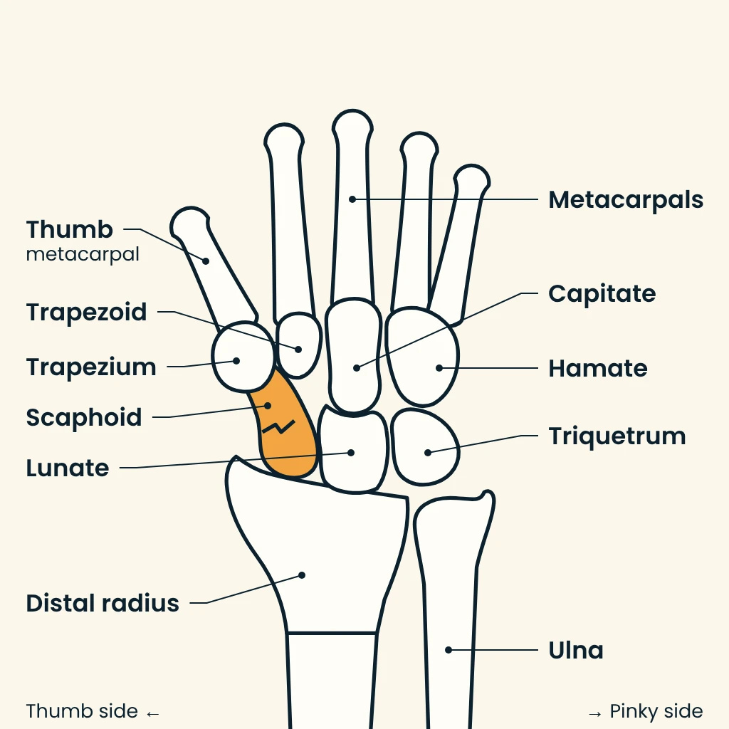
The scaphoid can articulate with all five surrounding bones, the distal radius, capitate, lunate, trapezium, and trapezoid, a 2011 review in the International Journal of Emergency Medicine notes.
The bone is easiest to locate with the thumb held in a thumbs-up position. It sits at the base of the hollow made by the thumb tendons on the back of the wrist, an area called the anatomic snuffbox, and that hollow is typically where tenderness or pain is felt most when the scaphoid breaks, per the AAOS.
Crash Forces on the Wrist
A scaphoid fracture usually happens when a person falls onto an outstretched hand and the weight lands on the palm, and the injury can also happen in motor vehicle collisions. In that kind of fall, the end of the radius may break as well, depending on the position of the hand on landing.
Scaphoid fractures are some of the most common broken bones, and any hard, sudden impact to the wrist can break the scaphoid, with car accidents among the common causes Cleveland Clinic lists.
The typical mechanism in the International Journal of Emergency Medicine review is a fall on the outstretched hand with the wrist in radial deviation, tilted toward the thumb, so the palm takes the impact. That load drives the scaphoid against the concave end of the radius, and the fracture is most likely to occur in the middle of the bone. The same mechanism also puts the back of the radius and the scapholunate ligament at risk, while falls on a wrist held in abduction carry an increased chance of a proximal pole fracture.
Scaphoid fractures account for 2% to 7% of all fractures and 82% to 89% of carpal fractures, according to that review, and they predominantly occur in young, active males. They also occur in people of all ages, including children, the AAOS notes. The page on car accident shoulder injuries covers the other end of the arm.
Symptoms Near the Base of the Thumb
Swelling and pain at or near the anatomic snuffbox and on the thumb side of the wrist are the usual signs, according to the AAOS. The pain may be severe when the thumb or wrist moves, or when the person tries to pinch, grasp, push, or pull something.
Pain, swelling, tenderness, being unable to move the wrist, bruising or discoloration, and a deformity or bump that is not usually on the wrist make up the symptom list from Cleveland Clinic.
Unless the wrist is deformed, a broken scaphoid may not be obvious to the injured person, and with some of these fractures the pain is not severe and may be mistaken for a wrist sprain, the AAOS cautions. Wrist pain that does not go away within a day of injury may be a sign of a fracture, and prompt treatment may help avoid potential complications.
No reliable clinical test confirms or rules out the diagnosis, the 2011 review states. Pain with pressure on the snuffbox, pain with pressure on the scaphoid tubercle, and pain with axial pressure on the first metacarpal bone each had a sensitivity of 100% in the studies the review cites, but their specificity was 9%, 30%, and 48%. Grip strength diminished by more than 50% compared with the other side increases the positive predictive value for a scaphoid fracture, by the review‘s account.
Scaphoid Fracture Warning Signs
- Swelling and pain on the thumb side of the wrist
- Tenderness in the anatomic snuffbox at the base of the thumb
- Pain when pinching, grasping, pushing, or pulling
- Pain that gets worse when moving the thumb or wrist
- Bruising or discoloration around the wrist
- Not being able to move the wrist
- A deformity or bump not usually on the wrist
- Wrist pain that does not go away within a day
Waist, Pole, and Displacement Patterns
A scaphoid fracture is usually described by its location within the bone. The mid-portion, called the waist, is where the scaphoid most commonly breaks, and fractures can also occur at the proximal and distal ends, the AAOS explains.
Each location has a name in the Cleveland Clinic breakdown: a distal pole fracture is closest to the hand and fingers, a waist fracture sits in the middle part of the bone and is the most common type, and a proximal pole fracture is at the end closest to the forearm. Under the Mayo classification described in the International Journal of Emergency Medicine review, 10% of fractures are proximal, 70% middle, and 20% distal.
Displacement is the second measure. In a non-displaced fracture the bone fragments line up correctly, while in a displaced fracture they have moved out of their normal position, sometimes leaving gaps between the pieces or overlapping, per the AAOS. Displaced fractures are much more likely to require surgery to repair, Cleveland Clinic says.
The Herbert classification sorts fractures by stability. A dislocation of more than 1 mm or an angulation of more than 15° between the fragments marks a fracture as unstable, and additional fractures, trans-scaphoid perilunate dislocations, multi-fragment fractures, and proximal pole fractures are also classified as unstable, according to the 2011 review.
Why Kentucky Hires
Sam Aguiar Injury Lawyers
Blood Supply That Runs Backward
In most bones of the arms and legs, blood runs from the part closest to the body toward the fingers or toes. The scaphoid works in reverse, the AAOS explains: blood enters the end closest to the thumb and runs back toward the end closest to the body. For that reason, fractures of the distal scaphoid heal much more quickly than fractures of the proximal scaphoid.
Eighty percent of the scaphoid consists of cartilage, leaving limited space for supplying arteries to enter, and the dorsal branch of the radial artery provides 75% of the blood supply, the International Journal of Emergency Medicine review reports. The proximal pole depends on blood reaching it from the distal pole through the bone, so a proximal fracture interrupts that route and makes healing of the proximal pole particularly difficult.
There is a risk that fractures of the proximal scaphoid will completely disrupt the blood supply and cause part of the bone to die, the AAOS warns. When a fragment loses enough blood flow, its cells die and the bone will not heal properly, a condition called avascular necrosis that can cause long-term problems in the wrist.
Displacement adds to the danger, since the blood supply may be disrupted especially in fractures whose fragments have moved, according to the AAOS. Because parts of the bone have a poor blood supply and a fracture can slow or stop that flow further, complications with healing are more common in scaphoid fractures than in fractures of other bones.
Sprain or Occult Fracture
A sprain is an injury to the ligaments that hold bones together, and 20 ligaments support the eight wrist bones, according to Cleveland Clinic. The scapholunate ligament, which connects the scaphoid and lunate bones, is the most commonly sprained one. Falls onto outstretched hands, sports injuries, and traumas like car accidents are the most common causes.
Wrist pain, swelling, bruising or discoloration, and trouble moving the wrist are the most common sprain symptoms on the Cleveland Clinic sprain page, a list that overlaps heavily with the signs of a broken scaphoid. Most wrist sprains can be treated at home with rest, ice, and over-the-counter medicine, and recovery usually takes a few weeks, Cleveland Clinic says.
Bone fractures are among the other injuries that most commonly happen alongside a wrist sprain, Cleveland Clinic notes, and a provider will probably use a wrist x-ray to check for bone damage. Some scaphoid fractures do not show up well, or at all, on x-rays, and Cleveland Clinic calls these occult scaphoid fractures.
The table below sets a wrist sprain beside non-displaced and displaced scaphoid fractures, drawing on Cleveland Clinic’s sprain and scaphoid fracture pages, the AAOS, and the 2011 review.
| Feature | Wrist Sprain | Non-Displaced Scaphoid Fracture | Displaced Scaphoid Fracture |
|---|---|---|---|
| What happens | Wrist ligaments stretched or torn | Bone broken; the fragments line up correctly | Fragments moved out of normal position, with gaps or overlap |
| Common signs | Wrist pain, swelling, bruising, trouble moving the wrist | Pain and swelling near the base of the thumb; some scaphoid fractures may be mistaken for a sprain | Pain, swelling, tenderness; sometimes a deformity or bump |
| First x-ray | Used to check the bones for damage | May look normal at first (occult fracture) | Displaced fractures are seen on radiographs |
| Further imaging | MRI or CT may be needed | MRI or CT, or a splint for 2 to 3 weeks and a repeat x-ray | CT can show whether the bones are displaced |
| Common treatment | Rest, ice, compression, elevation; sometimes a brace or splint | Cast or splint, often including the thumb | Much more likely to need surgery |
| Recovery range | Usually a few weeks | Around three months for most people; depends on severity and the treatments needed | Around three months for most people; depends on severity and the treatments needed |
Limits of Initial X-Rays
An x-ray is usually the first test, ordered to show whether the scaphoid is broken, whether the pieces are displaced, and whether other fractures or dislocations are present, according to the AAOS. In some cases the fracture does not show up right away, and scaphoid fractures often become visible on x-ray only after a period of time.
Initial radiographs detect at most 70% of all scaphoid fractures, according to the International Journal of Emergency Medicine review, which holds that anterior-posterior and lateral views should be standard and that at least two additional views are called for when a scaphoid fracture is suspected.
When a doctor suspects a fracture the film does not show, the AAOS describes wearing a wrist splint or cast for 2 to 3 weeks and returning for a follow-up x-ray, while avoiding activities that might cause further injury. The 2011 review is more skeptical of that repeat film: even on a repeated exam after 10 to 14 days, a scaphoid fracture is often missed because the added sensitivity is low.
Normal first films do not settle the question. Among patients with clinical signs of a scaphoid fracture and normal initial radiographs, approximately 20% do have a true fracture, a 2015 Cochrane review found. Because of the low healing potential of the scaphoid, its authors wrote, adequate diagnosis and treatment is vital to prevent complications such as nonunion.
MRI, CT, and Bone Scans
An MRI can sometimes show a scaphoid fracture before it can be seen on x-ray, and a CT scan can reveal the fracture and show whether the bones are displaced, the AAOS says, adding that doctors may use CT findings to help plan treatment. A provider might need an MRI or CT in addition to x-rays, Cleveland Clinic notes.
The Cochrane review pooled 11 studies of patients whose first radiographs were normal. The reviewers reported summary sensitivity and specificity of 0.72 and 0.99 for CT, 0.88 and 1.00 for MRI, and 0.99 and 0.86 for bone scintigraphy, a nuclear bone scan. CT and MRI had comparable diagnostic accuracy.
Bone scanning caught the most fractures but treated more wrists that were not broken. In a cohort of 1,000 patients, the reviewers estimated, 112 would be over-treated with a bone scan against 8 with CT, while CT would miss 56 fractures and a bone scan 2. A bone scan also carries radiation exposure and a diagnostic delay of at least 72 hours, the Cochrane authors note.
Timing changes MRI results too. Early MRI within 1 day after trauma has a limited sensitivity of 80%, while MRI performed after 19 days showed better results than bone scintigraphy, according to the 2011 review. When adequately performed, MRI also allows diagnosis of soft tissue and ligament injuries at the same time. The page on radiology imaging after a crash covers other scans used after a collision.
Casting and Thumb Spica Immobilization
Treatment depends on the location of the break, whether the fragments are displaced, how long ago the injury happened, and the patient’s age and general health, according to the AAOS. Non-displaced fractures closer to the thumb, at the distal pole, are likely to heal with proper protection and restricted activity, because that part of the bone has a good blood supply.
For those breaks, the cast or splint will usually sit below the elbow and include the thumb, and some fractures are held in a thumb spica cast covering the forearm, wrist, and thumb. Healing can be more difficult for a non-displaced break at the waist or proximal pole, the AAOS notes. A cast for that fracture may include the thumb and may extend above the elbow, and it typically stays on longer than one for a distal fracture.
Nearly every motion of the hand, wrist, and forearm moves the scaphoid and puts pressure on the fracture line, which makes the bone hard to immobilize, the International Journal of Emergency Medicine review points out. Generally, 6 weeks in a cast should be sufficient for most non-displaced and stable fractures, and the review found no study proving better healing with one cast type over another, with or without the thumb. Inadequate immobilization increases the chances of pseudo-arthrosis, a nonunion, by 30%, the authors report.
In some cases a doctor may recommend a bone stimulator, a small device that delivers low-intensity ultrasonic or pulsed electromagnetic waves to stimulate healing, the AAOS notes. If the bone has shifted, Cleveland Clinic describes a closed reduction, in which the provider realigns the bone by pushing and pulling the hand and wrist under anesthetic.
Screws, Grafts, and Surgical Repair
Surgery may be recommended when the scaphoid is broken at the waist or proximal pole, when the fracture is displaced, or both, with the goal of realigning and stabilizing the fracture so it has a better chance to heal, according to the AAOS. During reduction the surgeon moves the bone back into position, sometimes through a small incision, sometimes through an open incision, and sometimes with the help of a tiny camera called an arthroscope.
Metal implants, including screws, plates, or wires, then hold the scaphoid in place until the bone is fully healed. The incision may be on the palm side or the back of the wrist, depending on which part of the bone is broken. A bone graft, taken from the forearm, elbow, hip, or another area or supplied as a manufactured graft, may be used with or without internal fixation to stimulate bone production and healing, the AAOS writes.
Severe breaks may need open reduction and internal fixation, and a follow-up procedure may be needed to remove the pins, wires, or screws after healing, Cleveland Clinic says. Cleveland Clinic describes a graft as rare, used if the fracture is severely displaced or the scaphoid does not heal correctly.
Surgical treatment of non-displaced fractures has increased with improved, minimally invasive techniques, the 2011 review reports, because percutaneous screw fixation allows early functional treatment and can prevent an immobilization period of often 8 to 12 weeks. The authors note there is still insufficient evidence about which treatment is preferable for the non-displaced fracture. For specific fracture patterns, the AAOS describes an incision less than 1/2 inch long, with a special x-ray machine directing placement of the screw.
Recovery Time and Activity Limits
Most people need around three months to recover from a scaphoid fracture, Cleveland Clinic says, and the timeline depends on the fracture’s severity and the treatments needed. Physical therapy will probably be needed to regain strength and mobility in the wrist.
Unlike most other fractures, scaphoid fractures tend to heal slowly, and a cast or splint may be required for up to 6 months or until the fracture has healed, whether treatment was surgical or not, according to the AAOS. Unless a doctor approves more, the AAOS list of activities to avoid during that time includes lifting, carrying, pushing, or pulling more than 1 pound, throwing with the injured arm, contact sports, climbing ladders, and using machines that vibrate. Smoking can delay or prevent fracture healing, the AAOS notes.
Wrist stiffness after a scaphoid fracture is more common in patients who wear a cast for a long time or need more extensive surgery, according to the AAOS. Maintaining full finger motion matters throughout recovery, and a hand therapist may work on range of motion and strength. Even with hand therapy and a great deal of effort at home, some patients may not recover the motion and strength they had before the injury.
Wrist stiffness and reduced wrist strength were less frequently observed after a successful operation, according to the International Journal of Emergency Medicine review, which links the shift toward surgery partly to the demand for early return to productivity among young patients.
Nonunion, Necrosis, and SNAC Arthritis
A bone that fails to heal is called a nonunion, and nonunions are more common after scaphoid fractures because the blood supply to the bone is poor, the AAOS says. The reported incidence of scaphoid nonunion is between 5% and 15%, according to the 2011 review. Even when scaphoid fractures are adequately treated with casts, the reported nonunion rate has been as high as approximately 10%, a 2018 review in the Journal of Wrist Surgery reports.
Established nonunion typically leads to carpal collapse and osteoarthritis of the wrist, known as scaphoid nonunion advanced collapse, or SNAC, the Journal of Wrist Surgery review explains. Most scaphoid nonunions are symptomatic, but some cases are less symptomatic with gradual collapse and do not receive the required medical attention for a prolonged period. When the fracture lies distal to the apex of the scaphoid’s dorsal ridge, untreated breaks normally show a humpback deformity relatively early after the injury.
Over time, nonunion and avascular necrosis can lead to arthritis of the wrist, in which the joint cartilage becomes worn and frayed, sometimes until bone rubs on bone, per the AAOS. Aching, stiffness, decreased range of motion, and pain with lifting, gripping, or weightbearing are among its symptoms. Cleveland Clinic also lists compartment syndrome and malunion among the complications, especially when a fracture is not treated right away.
A symptomatic nonunion is best treated with surgery that includes debridement, realignment, and a vascularized or non-vascularized bone graft, with a success rate between 74% and 94% and much worse results for proximal nonunions, the International Journal of Emergency Medicine review reports. Once arthrosis develops, a lasting reduction of pain and functional improvement are often no longer achieved, and the review lists styloidectomy, denervation of the carpal joint, and removal of the scaphoid with a four-quadrant fusion as the rescue operations.
Medical Records Across the Recovery
The first exam sets the baseline. The AAOS describes a doctor asking how the injury happened, checking for tenderness directly over the scaphoid in the anatomic snuffbox, and looking for swelling, bruising, and loss of motion. Cleveland Clinic advises telling the provider what the person was doing before the symptoms started.
Each imaging report then adds detail: which part of the bone broke, whether the pieces are displaced, and, when the first film was clear, which later study found the break. Follow-up x-rays show whether the scaphoid is healing back together as it should, according to Cleveland Clinic, and the AAOS says doctors monitor healing with x-rays or other imaging studies. Operative notes and hand therapy records fill in the months between.
A dated log of each splint or cast change, each repeat film, and each therapy visit, with the motion and grip numbers recorded there, gives later providers a timeline to check against. Kept beside the imaging reports, that log puts the whole course of a scaphoid injury in one place.
Wrist Pain That Never Went Away?
A broken scaphoid can mean a clear first x-ray, weeks in a thumb spica cast, a screw or a bone graft, and stiffness or arthritis that shows up much later, and an insurer may judge the whole injury by the first emergency room note. Sam Aguiar Injury Lawyers gathers the emergency and imaging records, the casting and operative notes, and the hand therapy records so each stage of your recovery is on paper. Under our Bigger Share Guarantee®, you always walk away with more than the firm after bills, liens, and case costs. Start with a free case review.