

Car Accident Facial Fractures
Key Takeaways
- Nasal bone fractures are the most common type of facial fracture, representing 40% to 50% of cases.
- Motor vehicle accidents account for 50% of Le Fort midface fractures described in the literature, with higher speed collisions tending to produce higher grade patterns.
- After facial trauma, more surgery may be needed in 6 to 12 months to correct changes in appearance.
Facial Fracture Basics
A facial fracture is a broken bone in the face. These breaks fall under facial trauma, also called maxillofacial trauma, and car accidents and falls are among the causes. Facial bones are thinner than a lot of other bones in the body, and that makes them prone to injury.
The most common facial fractures involve the nose, the forehead, the eye sockets, the cheekbones, and the upper or lower jaw, along with the tripod fracture that takes in the eye socket, cheekbone, and upper jaw together and the midface breaks called Le Fort fractures. Facial injuries can affect the upper jaw, lower jaw, cheek, nose, eye socket, or forehead, and car and motorcycle accidents are among the common causes.
Across all causes, physical assault comes first, followed by vehicular accidents and falls. The majority of facial fractures are not life threatening, though some can lead to serious problems with vision, the airway, breathing, or the central nervous system.
Collision Forces and Bone Strength
Some parts of the facial skeleton take far more force to break than others. The outer rim of the eye socket is made of very thick bone, so it takes a lot of force to injure, and car accidents are the most common cause of orbital rim fractures. A person with an orbital rim fracture likely has other facial fractures as well.

Motor vehicle accidents are the most common cause of Le Fort fractures described in the literature, accounting for 50% of these injuries, with higher speed collisions tending to produce higher grade fracture patterns. Lower energy events such as falls from standing are more likely to result in lower grade Le Fort fractures.
A patient in a motor vehicle accident usually suffers multiple or compound mandibular fractures, while a jaw broken in an altercation is more likely to be single, simple, and nondisplaced. In automobile accidents, the condyle at the top of the lower jawbone was the most common fracture site.
Symptoms After a Blow to the Face
General symptoms include bruising, swelling, pain or tenderness, facial numbness, and disfigurement. A broken nose may add nosebleeds and difficulty breathing through the nose. Fractured eye sockets may bring blurred or double vision, sunken or bulging eyeballs, and difficulty moving the eyes. A fractured jaw may cause drooling, difficulty chewing or speaking, pain when opening the mouth, and loose, broken, or missing teeth.
A physical exam may show bleeding from the nose, eyes, or mouth and upper and lower teeth that no longer line up. Abnormal feeling on the cheek, irregularities of the face that can be felt by touch, and movement of the upper jaw while the head is still may suggest broken bones.
Swelling can hide the shape of the injury at first. A misshapen nose may not be apparent until the swelling goes down. The signs below are reasons to go to the nearest emergency room.
Facial Fracture Emergency Signs
- Open wounds where bone is visible
- Bloody or clear discharge from the nose
- Blurred or double vision, or problems moving the eyes
- Trouble swallowing or breathing
- A displaced jaw or nose
- Upper and lower jaws that do not meet properly, or pain when moving the jaw
- Loose teeth
- Pain and swelling in the face
Broken Nose and Septal Injury
Nasal bone fractures are the most common type of facial fracture, representing 40% to 50% of cases. The nose sticks out from the plane of the face and sits at its center, and that position predisposes it to injury. When high velocity trauma breaks the nose, other facial fractures are more likely to occur at the same time.
A fractured nose often occurs with other fractures of the face, and nose and neck injuries are often seen together, since a blow forceful enough to injure the nose may be hard enough to injure the neck. Damage to the cartilage can cause a collection of blood to form inside the nose. If that blood is not drained right away, it can cause an abscess or a permanent deformity that blocks the nose.
Not every broken nose needs to be reset. No further intervention is needed if there is no deformity or the patient is unconcerned about the appearance. When the bones have shifted, a doctor may be able to return them to their normal position within the first 2 weeks after the break.
Why Kentucky Hires
Sam Aguiar Injury Lawyers
Orbital Floor and Blowout Fractures
The orbit is the bony socket that holds the eye. A blowout fracture, a break along the floor or thin inner wall of that socket, is the most common type of orbital fracture, and car accidents and falls often cause orbital floor fractures.
The orbital bones are thin and delicate, which leaves them prone to fractures from relatively minor trauma. A blowout fracture may let the contents of the orbit herniate or become trapped. A trapped eye muscle can cause limited eye movement, pain, double vision, optic nerve compression, and visual disturbances.
Double vision on upward gaze, limited upward gaze, tenderness or step-offs along the lower orbital rim, and swelling and bruising around the eye are findings characteristic of orbital floor fractures that warrant further imaging. In adults, a classic blowout fracture shows visible soft tissue changes such as a sunken eye, called enophthalmos.
Lower Jaw Breaks and Bite Changes
The mandible, or lower jaw, is a mobile, ring-like bone that frequently fractures in more than one location. Because of that ring shape, multiple fractures are seen in more than 50% of cases. Broken jawbones are the second most common facial fracture.
Pain in front of the ear that gets worse with movement is one symptom of a broken or dislocated jaw. Others include difficulty chewing, misaligned teeth or jaw, difficulty opening the mouth widely, loose or damaged teeth, and numbness of the face, particularly the lower lip. Jaw joint pain is among the possible complications of a TMJ and jaw injury.
Cheekbone and Zygomatic Complex Injuries
The zygoma, or cheekbone, meets the surrounding bones at four points, and together they form the zygomaticomaxillary complex, or ZMC. Estimates suggest ZMC fractures account for 25% of facial fractures. They are sometimes called tripod fractures, though tetrapod fracture is the correct term given the four connections.
One of those connections crosses the lower rim of the eye socket, so orbital floor fractures frequently occur alongside ZMC fractures. Altered sensation in the face is a common result, because sensory nerves run close to the cheekbone. One of them, the infraorbital nerve, carries feeling from the cheek, upper lip, nose, and upper front teeth.
A pushed-in cheekbone may cause facial flattening, though swelling can hide that flattening early on. A depressed fracture of the zygomatic arch may also impede movement of the temporalis muscle and cause trismus, a limit on how far the mouth opens.
Le Fort Midface Patterns
Le Fort fractures are named for René Le Fort, a French military surgeon who studied midface injury patterns in 1901. They sort into three types, all of which separate the upper jaw from the skull. Type I runs horizontally through the upper jaw above the roots of the teeth, separating the hard palate from the upper face. Type II is a pyramid that takes in the nasal bridge, upper jaw, orbital floor, and lower orbital rim. Type III separates the entire midfacial skeleton from the base of the skull.
Le Fort fractures often occur asymmetrically, and more than one type can coexist on the same side of the face. They seldom occur in isolation, often arriving with other injuries that are sometimes life threatening. Type III fractures may come with a cerebrospinal fluid leak from an injury to the skull base, and the risk of life threatening bleeding in type II and III injuries is higher than with other facial injuries.
Four Fracture Types Side by Side
The table lines up four common patterns: nasal, orbital floor, mandible, and Le Fort fractures.
| Feature | Nasal | Orbital Floor | Lower Jaw | Le Fort |
|---|---|---|---|---|
| Accident link | Motor vehicle accidents are among the typical causes | Car accidents and falls often cause them | Condyle is the most common site in automobile accidents | Motor vehicle accidents account for 50% |
| Common signs | Nosebleed, bruising around the eyes, trouble breathing through the nose | Double vision and limited upward gaze, sunken eye | Misaligned teeth, trouble opening the mouth, lower lip numbness | Midface instability, open bite, reduced midface feeling |
| Usual imaging | Often none; CT for head, skull base, or complex injuries | CT, the imaging method of choice | Mandibular x-ray series, panoramic x-ray, or CT | Fine-cut CT |
| Typical treatment | Reset not always needed; bones may be moved back within 2 weeks | Nondisplaced breaks may be treated without surgery; repair generally within 14 days | Jaws wired together, or plates and screws | Surgery a few days after injury, ideally within a week |
CT Scans and the Imaging Workup
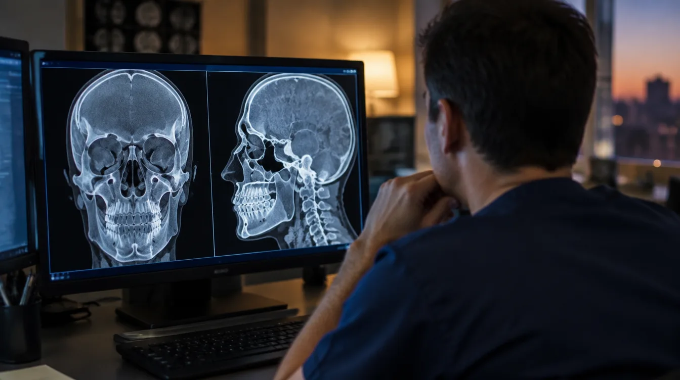
Computed tomography (CT) is the most common imaging test used to diagnose facial fractures. For midface trauma, fine-cut CT, with slices of 3 mm or less, is the essential part of the evaluation, particularly because patients are often unable to cooperate with a physical exam after multiple injuries. Three-dimensional reconstructions show the surgeon and the patient how far the injuries extend.
Imaging for an isolated nasal fracture is rarely needed, and the diagnosis is typically made by clinical observation alone, with CT scans performed for suspected head injuries, skull base fractures, or complex facial injuries. For the lower jaw, a mandibular x-ray series, a panoramic dental x-ray, and CT are used, and CT is indicated when other facial fractures are suspected. CT is the imaging method of choice for orbital fractures.
CT imaging is also excellent for finding related injuries in the cervical spine, skull, and face, and skull fracture (40.7%) is among the common injuries that accompany complex maxillofacial trauma.
Reduction, Fixation, and Surgery
Treatment centers on two steps: reduction, which resets the broken bones in their correct positions, and fixation, which keeps them there long enough to heal and sometimes requires surgical plates, screws, and wires. Closed reduction resets the bones without incisions. Open reduction is surgery, used when the fractures are too complex to reset by hand.
Surgery is done if the injury prevents normal functioning or causes a major deformity. Treatment goals include a clear airway, control of bleeding, fixing broken bone segments, preventing scars where possible, and preventing long-term double vision or sunken eyes or cheekbones.
Orbital floor surgery generally must be performed within 14 days of injury to prevent fibrosis, and most surgeons wait 24 to 72 hours for swelling to subside. Midface reconstruction for Le Fort fractures is typically scheduled a few days after the injury and ideally done before a week has passed. For the lower jaw, jaw wires are usually left in place for 6 to 8 weeks, while many surgeons now use rigid wiring for 2 weeks or less before switching to elastic bands, and plates and screws are often used when long wiring is not practical.
Recovery Time Ranges
Recovery is different for everyone and depends on the extent of the trauma, the treatment, and the body’s own healing. After facial reconstructive surgery, swelling and bruising usually fade after about two weeks, but complete recovery can take up to a few months.
The bruised look of a broken nose most often disappears after 2 weeks. A mild jaw fracture may mean a liquid diet for four to six weeks, and after jaw surgery it may be a month or more before the bone heals completely, and it may be weeks or months before usual activities resume.
After orbital fracture surgery, most people can return to school or work within a week, though strenuous activity, air travel, and deep-sea diving may be limited for several weeks. Some people develop nerve pain that can last up to nine months.
Lasting Complications and Revision Surgery
Some people may have no long-term effects after a facial fracture, but long-term complications are possible, including sinus problems, breathing problems, a change in taste or smell, tingling or numbness, vision problems, headaches, and tenderness in the head or face. Some facial fractures can also cause a cerebrospinal fluid leak.
In the lower jaw, malocclusion, a bite that no longer fits together, is the most common complication of mandibular fractures and their treatment. Reduced feeling in the lower lip and chin is also extremely common, with rates as high as 50%. Among patients treated with open reduction and internal fixation, only 7% describe long term complications such as abscess, bone that heals out of position or fails to heal, and exposed hardware.
Around the eye, persistent double vision could require another surgery, and enophthalmos may worsen with time. Persistent facial deformity and the need for revision surgery are Le Fort complications, and more surgery may be needed in 6 to 12 months to correct changes in appearance. Eye involvement is not always severe: in a survey of 40 patients treated at a level 1 trauma center for motor vehicle accident eye injuries, most reported no change in vision and returned to work and driving within a year.
Medical Records That Track the Injury
Visual acuity, eye muscle function, double vision, the degree of enophthalmos, and any abnormal sensation should be documented before an orbital fracture repair. For midface fractures, the bite is recorded with the Angle classification, and dental records, when available, serve as a reference for surgical reduction.
An otolaryngologist, plastic surgeon, or oral surgeon is the usual lead for Le Fort repairs, with a neurosurgeon, ophthalmologist, or dentist added when needed, and each visit leaves its own notes. Cuts, scarring, and other soft tissue damage to the face are separate facial injuries.
Photos of the face taken in the first days after an accident, and again as the swelling settles, show changes that a chart can only describe in words. Keeping the CT reports, operative notes, and dental and eye records together puts the whole repair history in one place.
Facial Fracture Still Healing After Surgery?
A face can look healed long before the numbness, the bite, or a planned revision surgery has settled, and an insurer may judge the injury from the early notes. Sam Aguiar Injury Lawyers tracks each stage of care, from the first CT to any revision surgery, so the record shows the whole recovery. Under our Bigger Share Guarantee®, you always walk away with more than the firm after bills, liens, and case costs. Start with a free case review.