

Radiology Imaging After a Car Accident
Key Takeaways
- Bone x-rays give little information about muscles, tendons, or joints, so a clean x-ray does not rule out a ligament tear or disc injury.
- In surgically confirmed subtle Lisfranc injuries, x-ray found the injury in 48.4% of patients, CT in 87.1%, and MRI in 96.8%.
- MRI uses magnets and radio waves with no ionizing radiation and usually takes about 30 to 60 minutes.
Radiology Basics After a Crash
Radiology is the branch of medicine that uses imaging to look inside the body. After a car accident, the usual tests are x-rays, computed tomography (CT), magnetic resonance imaging (MRI), and ultrasound. Each one sees different tissue, so a test that is normal for one kind of injury can say little about another.
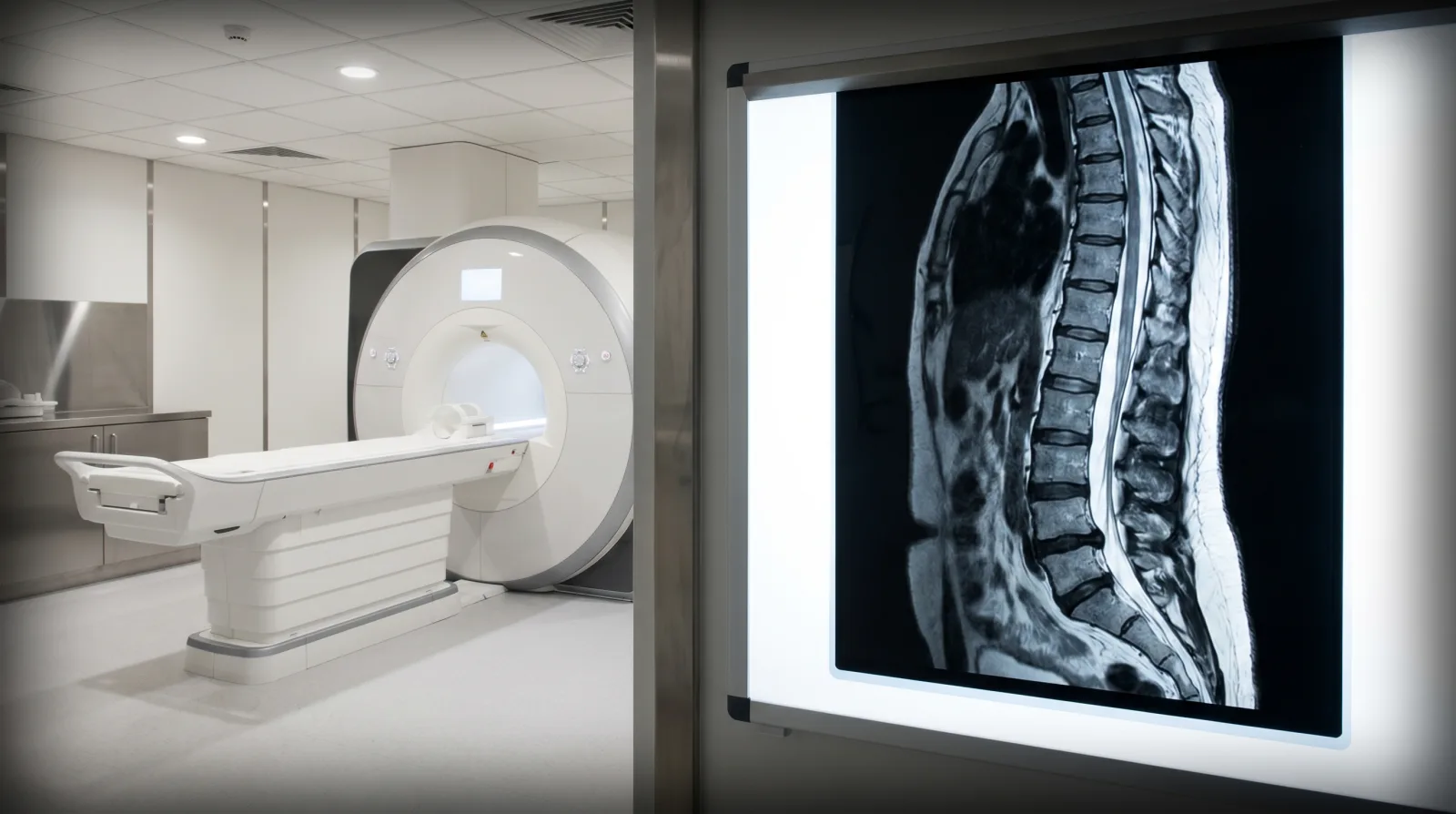
A radiologist reads the images. The radiologist is the doctor who supervises the exam, interprets the images, and writes a report for the provider who ordered it.
First-Line X-Rays in the Emergency Room
An x-ray is usually the first picture taken after an accident. Dense bone absorbs much of the radiation, so bone shows up white, soft tissue appears in shades of gray, and air appears black. That contrast makes x-rays good at showing broken bones and dislocated joints.
The same page names the limit plainly: x-ray images are among the clearest views of bone, but they give little information about muscles, tendons, or joints. A clean x-ray rules out some fractures. It does not rule out a torn ligament or a damaged disc.
Computed Tomography for Trauma
A CT scan also uses x-rays, but a computer turns them into cross-sectional images called slices. A CT scan creates detailed pictures of the brain, chest, spine, and abdomen, and most complete scans take only a few minutes.
Speed and bone detail are why CT is common in trauma care. CT can image complicated fractures, subtle fractures, and dislocations, and for a suspected spine injury, 3-D reconstructed images can be made without extra radiation exposure. CT still uses ionizing radiation, which is among its risks.
Symptoms That Lead to More Testing
Imaging decisions follow symptoms and the physical exam. MRI helps diagnose or rule out acute spinal cord compression when an exam shows muscle weakness or paralysis. Follow-up imaging is used when a doctor suspects a fracture that the first x-ray does not show.
Head injuries carry their own warning list. The people checking on someone after a head injury should call 9-1-1 or go to an emergency department right away for any of the danger signs below.
Head Injury Danger Signs
- A headache that gets worse and does not go away
- Weakness, numbness, decreased coordination, convulsions, or seizures
- Repeated vomiting
- Slurred speech or unusual behavior
- One pupil larger than the other
- Confusion, restlessness, or agitation, or not recognizing people or places
- Loss of consciousness, heavy drowsiness, or being unable to wake up
Why Kentucky Hires
Sam Aguiar Injury Lawyers
Magnetic Resonance Imaging for Soft Tissue
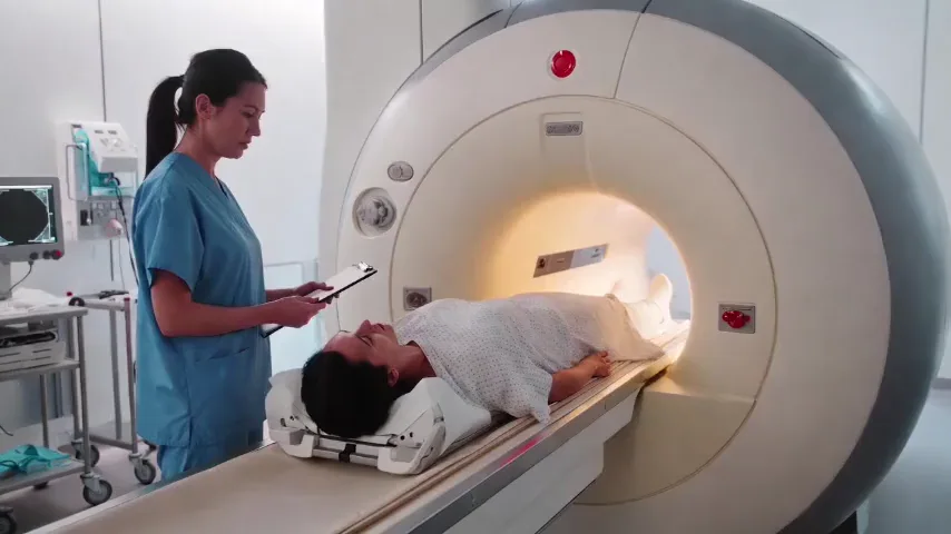
MRI works differently. It uses powerful magnets and radio waves to create pictures of the body, with no ionizing radiation. The scan usually lasts about 30 to 60 minutes, and some exams use a contrast dye given through a vein so the radiologist can see certain areas more clearly.
MRI shows the structures an x-ray cannot. MRI may be more useful for meniscus and ligament tears in the knee, rotator cuff and labrum tears in the shoulder, and the spine, where both the bones and the spinal cord can be evaluated. MRI can find subtle or hidden fractures and bone bruises that do not appear on x-ray.
For the neck and back, MRI is commonly used for trauma to the bone, disc, ligament, or spinal cord, and it is the best available method to evaluate ligament injuries.
Three Common Scans Side by Side
The table lines up the three tests: x-ray, CT, and MRI.
| Feature | X-Ray | CT Scan | MRI |
|---|---|---|---|
| How it works | X-rays pass through the body | X-rays turned into cross-sectional slices | Magnets and radio waves |
| Shows best | Broken bones and dislocations | Complicated and subtle fractures | Ligaments, tendons, discs, spinal cord, bone bruises |
| Main limit | Little detail on muscles, tendons, or joints | MRI is the better test for ligament injuries | Longer exam, about 30 to 60 minutes |
| Ionizing radiation | Yes | Yes | No |
Fractures and Tears Plain Films Miss
Some injuries are known for hiding on a first x-ray. Among patients with surgically confirmed subtle Lisfranc injuries, a midfoot injury, x-ray reached the correct diagnosis in 48.4% of patients, CT in 87.1%, and MRI in 96.8%. Non-weight-bearing x-rays had poor accuracy for these injuries.
Wrist fractures follow a similar pattern: a scaphoid fracture sometimes does not show up on an x-ray right away, it often becomes visible only after a period of time, and an MRI can sometimes show the fracture first.
Chest injuries can hide too. CT found chest injuries in 39% of patients whose first chest x-ray was normal and who had a severe mechanism of injury.
Ultrasound and Arthrography
Ultrasound uses sound waves instead of radiation. Musculoskeletal ultrasound produces pictures of muscles, tendons, ligaments, nerves, and joints, and its common uses include rotator cuff tears and ligament sprains or tears.
Arthrography adds contrast to a joint before imaging. In direct arthrography, a radiologist injects contrast straight into the joint, often using fluoroscopy or ultrasound to place the needle, and the images may then be taken with MRI or CT. The method is particularly effective for problems with ligaments, the labrum, tendons, and cartilage, especially in the shoulder after a dislocation and in the hip, wrist, and elbow.
Brain Imaging After a Concussion
A normal head scan does not rule out a concussion. A brain scan such as a CT is not needed to spot a mild traumatic brain injury or concussion, though it may sometimes be used for patients at risk for bleeding on the brain.
Diffusion tensor imaging (DTI) is a newer MRI method that maps the brain’s white matter. There is no relevant literature supporting DTI in the initial imaging evaluation of acute head trauma.
Age-Related Findings on Spine Scans
Spine MRIs often show wear that was there before an accident. Among 3,110 people who had no back pain, disc degeneration appeared in 37% of 20-year-olds and 96% of 80-year-olds, and disc bulges rose from 30% to 84% across the same ages.
Many of these findings are likely part of normal aging and unrelated to pain. That is why a radiologist’s description of degeneration says little on its own about whether a new injury happened. Symptoms, their timing, and comparison with earlier scans are what show whether an injury is new.
Reading the Radiology Report
Every study ends in a written report. The usual sections are the history or reason for the exam, any comparison with prior studies, the technique used, the findings, and the impression.
The impression is where the radiologist summarizes the most important findings and possible causes, and it is the most important part of the report for the patient and the doctor. It may recommend more imaging, a follow-up study after a period of time, or comparison with outside scans the radiologist did not have. A finding marked “unremarkable” means the radiologist saw nothing concerning in that area, a term that appears often in spine and joint MRI reports.
Timing and Follow-Up Studies
A first x-ray in the emergency room is not always the last study. For scaphoid fractures, a splint or cast for 2 to 3 weeks and a follow-up x-ray are one way to catch a fracture the first film missed.
Radiologists compare a new exam with available prior exams and list those studies in the report. A clean baseline from before the accident, or an early scan right after it, gives the next radiologist something to measure against.
Imaging Records for an Injury Case
Each report and image set becomes part of the medical record. The ordering provider’s note explains why the test was done, the radiologist’s report states what was seen, and the images themselves can be reviewed again by another physician.
Copies of both the reports and the images are worth requesting from each facility, since outside radiologists often cannot see studies done elsewhere. Gaps between the first x-ray and a later MRI are easier to explain when the symptoms that drove each order are written down along the way.
A normal first x-ray is one data point in the record, not the whole picture. Keeping the emergency films, any later MRI or CT, and each radiology report together puts the full imaging history in one place.
X-Ray Came Back Clean, but the Pain Stayed?
A normal x-ray can leave you hurting with no answer, and an insurer may treat that first film as the last word on your injury. Sam Aguiar Injury Lawyers pulls together the emergency records, the later MRI or CT, and each radiology report so the injury on paper matches the injury you live with. Under our Bigger Share Guarantee®, you always walk away with more than the firm after bills, liens, and case costs. Start with a free case review.