

Car Accident Lower Leg Injuries
Key Takeaways
- Most tibial shaft fractures take 4 to 6 months to heal completely, according to the American Academy of Orthopaedic Surgeons, and some take even longer, especially open fractures or those broken into several pieces.
- The floor or a foot control was the source of an estimated 93 percent of moderate or more severe lower leg injuries among belted drivers in frontal crashes, a 2012 analysis by the National Highway Traffic Safety Administration found.
- Acute compartment syndrome, one of the most time-sensitive orthopedic emergencies, has reported incidences of 2 to 9% in tibial fractures depending on fracture type and severity, according to a 2025 meta-analysis in BMC Musculoskeletal Disorders.
Tibia and Fibula Anatomy
The American Academy of Orthopaedic Surgeons (AAOS) describes the lower leg as two bones, the tibia and the fibula. The tibia, or shinbone, is the larger of the two. It supports most of a person’s weight and is an important part of both the knee joint and the ankle joint.
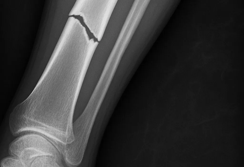
The fibula, the calf bone, does not support body weight the way the shinbone does, so a person with a broken fibula may still be able to put weight on the injured leg, Cleveland Clinic notes. With a broken tibia, a person probably will not be able to stand, walk, or put weight on the leg, Cleveland Clinic says.
At the top, the tibia widens to help form the knee joint. The AAOS calls this upper portion the proximal tibia and notes that the part supporting the joint is sometimes called the plateau. Its top surface is cancellous bone, which has a honeycombed appearance and is softer than the thicker bone lower in the tibia.
Between the knee and the ankle runs the tibial shaft. The tibia is the most commonly fractured long bone in the body, and for classifying breaks the AAOS divides the shaft into thirds: distal, middle, and proximal.
Footwell Intrusion in Frontal Crashes
A 2012 analysis by the National Highway Traffic Safety Administration (NHTSA) found that the floor or a foot control was the source of an estimated 93 percent of moderate or more severe lower leg injuries among belted drivers in frontal crashes. NHTSA estimated that the floor or toe pan pushed into the driver’s seating position in 2 percent of all vehicles in frontal crashes, and most often, at an estimated 6 percent, in a left side small overlap impact.
When that intrusion happened, the odds of a lower leg injury were twice the odds without it across all frontal crashes, the NHTSA report found, and the odds ratio rose to seven in full frontal, left offset, and small overlap crashes. The report estimated that each additional centimeter of intrusion increased the odds of lower leg injury by 14 percent in full frontal crashes and 5 percent in left offset and small overlap crashes.
Knee to dashboard impact from forward motion, and intrusion into the passenger compartment, have both been identified as significant mechanisms of injury to the lower leg, a 2018 review in African Health Sciences reports. Trapping of the knee or the foot may produce a twisting force on the tibia and fibula, the authors write, and combined with the forces from forward motion and intrusion, that could fracture both bones.
Several studies have reported more injuries below the knee in shorter female occupants, the African Health Sciences review notes, and data from the Crash Injury Research and Engineering Network identified short stature as a positive predictor of tibial fracture. A figure in the review explains that a shorter driver may need to extend the knee to reach the pedal.
Early Signs of a Broken Shin
A tibial shaft fracture usually causes immediate, severe pain, the AAOS notes. Other symptoms may include being unable to walk or bear weight on the leg, deformity or instability of the leg, bone tenting the skin at the fracture site or protruding through a break in the skin, and occasional loss of feeling in the foot.
With a fracture just below the knee, the AAOS lists pain that is worse when weight is placed on the leg, swelling around the knee with limited bending, and a knee that may look out of place. A pale appearance or cool feeling in the foot may suggest that blood flow to the foot is impaired, and numbness or pins and needles around the foot raises concern about nerve injury or excessive swelling within the leg.
Pain, tenderness, swelling, bruising or skin discoloration, a bump or deformity, and being unable to move the leg are among the symptoms of a broken fibula that Cleveland Clinic lists.
Pain more intense than what would be expected from the injury itself, which grows worse when the involved muscles are used or stretched, is the classic sign of acute compartment syndrome, according to the AAOS.
Lower Leg Warning Signs
- Immediate, severe pain in the shin
- Inability to walk or bear weight on the leg
- A leg that looks deformed, twisted, or shortened
- Bone tenting the skin or showing through a wound
- Swelling around the knee with limited bending
- A pale or cool foot
- Numbness or pins and needles around the foot
- Pain far worse than the injury seems to explain
Tibial Plateau Fractures Below the Knee
Fractures of the tibial plateau occur when a force drives the lower end of the thighbone into the soft bone of the plateau, similar to a die punch, the AAOS explains. The impact often compresses the cancellous bone and leaves it sunken. That damage to the joint surface may result in improper limb alignment and, over time, may contribute to pain, arthritis, instability, and loss of motion.
Young people often sustain these fractures in high-energy injuries such as motor vehicle accidents, falls from a considerable height, and sports trauma, while older people with poorer quality bone often sustain them after a low-energy injury, such as a fall from a standing position, according to the AAOS. Skin, muscle, nerves, blood vessels, and ligaments may be injured at the same time, and the AAOS says the broken bone and any soft tissue injuries must be treated together.
In a 2022 study in the Journal of Orthopaedic Surgery and Research of 1,165 patients with tibial plateau fractures at five trauma clinics in Colombia, 95.7% of the fractures were caused by traffic accidents, most of them involving motorcycles, and 78% of patients had associated ligament and meniscal injuries. The Schatzker classification used in the study has six types, with each step up representing an increase in severity, and the study found type VI (23%) and type V (19.1%) the most common. The page on knee injuries after a car accident covers ligament and meniscus tears in more detail.
Why Kentucky Hires
Sam Aguiar Injury Lawyers
Shaft Breaks Along the Shin
It typically takes a major force to break the tibial shaft, and the AAOS names motor vehicle collisions as a common cause. In many tibia fractures the fibula is broken as well. A high-energy collision such as a car or motorcycle crash can break the bone into several pieces.
The AAOS sorts shaft fractures by pattern. A transverse fracture runs as a straight horizontal line across the shaft, and an oblique fracture has an angled line. A spiral fracture encircles the shaft like the stripes on a candy cane and is caused by a twisting force. A comminuted fracture leaves the bone in three or more pieces. The pieces may line up correctly, a stable fracture, or sit out of alignment, a displaced fracture.
Cleveland Clinic adds the segmental fracture, in which the bone breaks in at least two places and leaves a separate segment of bone between the breaks.
The ends of broken bones are often sharp and can cut or tear surrounding muscles, nerves, or blood vessels, the AAOS warns.
Fibula Fractures of the Calf Bone
Broken fibulas rarely happen on their own, Cleveland Clinic notes. It is much more common to break the calf bone along with the tibia, and fibula fractures usually happen along with other broken bones in the leg or ankle. They usually result from a serious fall or car accident.
A distal fibula fracture is closer to the ankle and a proximal fibula fracture is closer to the knee, by Cleveland Clinic‘s account. Immobilization in a splint or cast is the first treatment Cleveland Clinic lists, and some people may be able to wear a walking boot. A person with an isolated fibula fracture probably will not need surgery, Cleveland Clinic says. When surgery is needed, the surgeon realigns the pieces, secures them in place, and puts the leg in a cast.
Most fibula fractures heal completely in six to eight weeks, although more severe fractures can take longer, Cleveland Clinic reports. Fibula breaks usually heal faster than other leg fractures because the fibula does not support weight when a person stands and moves. The page on car accident foot and ankle injuries covers breaks at the ankle end of the leg.
Open Breaks Through the Skin
The AAOS defines an open fracture, also called a compound fracture, as one with an open wound or break in the skin near the broken bone, most often caused by a fragment of bone breaking through the skin at the moment of injury. Most open fractures come from a high-energy accident such as a motor vehicle collision, and these patients often have additional injuries to other parts of the body.
In many high-energy injuries the bone can be seen protruding through the wound. In other cases the wound may be no larger than a puncture, yet the AAOS says the damage to the muscles, tendons, nerves, veins, and arteries around the bone can still be extensive. For that reason, any serious fracture with an open wound in the area is considered an open fracture.
Once the skin is broken, bacteria from dirt and other contaminants can enter the wound, so early treatment focuses on preventing infection, the AAOS explains. In the emergency room, the AAOS says, antibiotics are given along with a tetanus booster if the patient has not had one within the last 5 years. Almost all open fractures are treated with surgery: debridement removes foreign and contaminated material and damaged tissue, and irrigation washes the wound.
Most severe open fractures are first stabilized with external fixation, the AAOS writes. In that method, pins or screws go through the skin into the bone above and below the break and attach to bars outside the leg. In most cases the fixator stays only until internal fixation is safe. Wounds too large to close may be sealed temporarily, often over antibiotic beads, and later covered with a skin graft, a local flap of nearby muscle, or a free flap of tissue moved from another part of the body.
In a 2018 study in the Indian Journal of Orthopaedics of 120 patients with Gustilo IIIB open tibial fractures, a grade associated with high rates of infection and nonunion, 64.7% of cases were the result of motor vehicle accidents. The infection rate in that study was 30.3%, and the authors concluded that these high-energy open fractures required an average of 8.5 months to heal.
Compartment Syndrome and Rising Pressure
The area between the knee and ankle has four major muscle compartments, the AAOS explains: anterior, lateral, superficial posterior, and deep posterior. Each is covered by a tough membrane called fascia that does not stretch or expand easily. When swelling or bleeding builds inside a compartment, pressure rises on the capillaries, nerves, and muscles, and blood flow to muscle and nerve cells is disrupted.
A 2025 meta-analysis in BMC Musculoskeletal Disorders notes that tibial fractures, particularly those from high-energy trauma, are the most common injuries associated with acute compartment syndrome, with reported incidences ranging from 2 to 9% depending on fracture type and severity. The authors identified younger adult age, male sex, high-energy trauma, and polytrauma as critical risk factors.
Severe pain, especially when the muscle within the compartment is stretched, is the classic sign, per the AAOS. Tingling or burning in the skin and a tight or full feeling in the muscle may also appear, while numbness or paralysis are late signs that usually indicate permanent tissue injury. With an acute injury, symptoms can become severe within a few hours, MedlinePlus notes.
Acute compartment syndrome is a surgical emergency with no effective nonsurgical treatment, the AAOS says. In the operation, called a fasciotomy, the surgeon cuts open the skin and fascia covering the affected compartment, and when swelling is severe the incision is repaired later, sometimes with a skin graft. If the diagnosis is delayed, permanent nerve injury and loss of muscle function can result, and MedlinePlus says permanent nerve injury can occur after less than 12 to 24 hours of compression.
Three Lower Leg Injuries Compared
The table sets three common crash injuries side by side, drawn from the AAOS and Cleveland Clinic pages cited above. They often overlap, since the AAOS notes that in many tibia fractures the fibula is broken as well.
| Feature | Tibial Plateau Fracture | Tibial Shaft Fracture | Fibula Fracture |
|---|---|---|---|
| Where it breaks | Top of the tibia, where it forms the knee joint | Along the length of the tibia, below the knee and above the ankle | The calf bone, closer to the knee or the ankle |
| Typical signs | Pain worse with weight, knee swelling, limited bending | Immediate severe pain, unable to bear weight, deformity | Pain, swelling, bruising; weight on the leg may still be possible |
| Imaging | X-rays and CT, MRI if x-rays are negative | X-rays, CT if more detail is needed | X-ray, CT, or MRI |
| Common treatment | Plates and screws when the joint is involved, sometimes bone graft | Intramedullary nail, plates and screws, or external fixation | Splint, cast, or walking boot; surgery for some |
| Recovery range | Full weightbearing may wait 3 months or more | Most heal in 4 to 6 months | Most heal in 6 to 8 weeks |
X-Rays, CT, and MRI Findings
The AAOS lists x-rays as the most common way to evaluate a fracture. They show whether the tibia is broken, the type of fracture and where it sits, whether the knee or ankle joint is involved, and whether the fibula is broken too. A CT scan shows a cross-sectional image and can, in some cases, detect subtle fractures whose lines are very thin and hard to see on an x-ray.
For a break just below the knee, doctors often use both x-rays and CT scans to determine the location and displacement of the fragments, the AAOS says. MRI is not a routine test for tibia fractures, but a doctor may order one to help determine whether there are additional injuries to the soft tissues around the knee. If a person has all the signs of a tibial plateau fracture while x-rays are negative, a doctor may order an MRI, because an injured bone often shows a reaction in the marrow that MRI can detect.
How the crash happened belongs in the chart too. For a broken leg from a car accident, the AAOS says it helps the doctor to know how fast the vehicle was going, whether the patient was the driver or a passenger, whether a seatbelt was worn, and whether the airbags went off, so the doctor can judge how the injury happened and whether the person may be hurt somewhere else. The page on reading an MRI report explains common report terms.
Nails, Plates, and External Fixators
Most surgeons currently use intramedullary nailing for tibia fractures, the AAOS reports. A specially designed metal rod, usually titanium, is inserted inside the tibia across the fracture, and screws at both ends keep the nail and bone in position during healing. Plates and screws attached to the outer surface of the bone are often used when nailing may not be possible, such as for fractures that extend into the knee or ankle joint.
Nonsurgical care may be recommended for closed fractures with very little movement of the fracture ends, or for patients who are poor candidates for surgery, the AAOS explains. A splint may come first, since it can be tightened or loosened to allow swelling to occur safely, and after several weeks a cast can be replaced with a functional brace.
When a plateau fracture pushes part of the joint surface down, the surgeon may need to lift the sunken fragments back into place, which leaves a hole in the supporting bone. The AAOS says that hole must be filled to keep the bone from collapsing, using a bone graft from the patient or a bone bank, or synthetic or natural products that stimulate healing. If the skin and muscle around the fracture are badly damaged, a temporary external fixator may hold the leg until surgery can be performed safely.
Cleveland Clinic notes that most people need internal fixation surgery to repair a tibia fracture, and that some need external fixation before it is safe to have that surgery.
Recovery Ranges by Fracture Type
Most tibial shaft fractures take 4 to 6 months to heal completely, according to the AAOS. Some take even longer, especially if the fracture was open or broken into several pieces or if the patient uses tobacco products. Cleveland Clinic gives the same four to six month range for most broken tibias and says more severe fractures take longer.
After a proximal tibia fracture, full weightbearing is usually discouraged until some healing has occurred, which may require as much as 3 months or more, according to the AAOS. Crutches, a walker, or a wheelchair fill that gap, sometimes with a knee brace. Once weight is allowed, the AAOS calls it very normal to feel weak, unsteady, and stiff.
Open fractures in the lower leg, where the tibia sits right under the skin, take longer to heal than fractures in the thigh or upper arm, the AAOS explains, and it is common to have stiffness, discomfort, and weakness that may improve for a year or more.
Muscle strength is likely to be lost in the injured area, so the AAOS describes physical therapy, often started while the patient is still in the hospital, to restore normal muscle strength, joint motion, and flexibility.
Nonunion, Infection, and Knee Arthritis
The AAOS defines a nonunion as a broken bone that fails to heal and a delayed union as one that takes longer than usual to heal. Nonunions are more likely after a high-energy injury such as a motor vehicle collision, because severe injuries often disrupt the blood supply to the broken bone. The tibia has a moderate blood supply, the AAOS notes, and a high-energy injury can damage the skin and muscle over the bone, destroying the external blood supply, and can destroy the internal supply in the marrow as well.
Patients with a nonunion usually feel pain at the site of the break long after the initial pain of the fracture disappears, and that pain may last months or even years, the AAOS says. Surgical options include bone grafting and exchange nailing, in which a previously inserted nail is removed and a larger one is inserted.
Infection is the most common complication of open fractures, according to the AAOS. It can occur early in healing or much later after both the wound and the fracture have healed, and a bone infection can become chronic (osteomyelitis) and lead to further surgeries. In the Indian Journal of Orthopaedics study, smoking was associated with an increased risk of infection, 65.4% against 24.7%.
Because a proximal tibia fracture may involve a weight-bearing joint, the AAOS lists loss of knee motion and stability, along with long-term arthritis, among the long-term concerns. The Journal of Orthopaedic Surgery and Research study cites earlier series that identified osteoarthritis after tibial plateau fractures in 73.34%, 26.4%, and 24% of cases.
Records That Track the Healing
The emergency exam sets the baseline. The AAOS describes a doctor looking for deformity, breaks in the skin, bruises, swelling, bony pieces pushing on the skin, and instability, then feeling along the tibia, leg, and foot and, if the patient is awake and alert, testing sensation and movement in the leg and foot.
Later entries follow the bone. After a proximal tibia fracture, doctors regularly schedule x-rays to see how well the fracture is healing, and with a brace or cast those films show whether the bone is changing position, according to the AAOS. A nonunion may be diagnosed when repeated imaging compared over several months shows a persistent gap or no progress in healing, per the AAOS. Operative reports, wound care notes from any fixator or flap, and physical therapy notes fill in the months between. The page on car accident femur fractures covers breaks higher in the leg.
A dated log of when crutches, a walker, or a boot were used, when weight on the leg was first allowed, and when each new symptom appeared gives later providers a timeline to check against. Kept with the discharge papers and therapy schedules, that log puts the full course of a lower leg injury in one place.
Shin Still Aching Months After the Crash?
A broken lower leg can mean an external fixator, a nail or plate, a skin graft, and months on crutches, and an insurer may judge the whole injury by the first emergency room visit. Sam Aguiar Injury Lawyers gathers the operative reports, the wound care notes, and the follow-up x-rays so each stage of your recovery is on paper. Under our Bigger Share Guarantee®, you always walk away with more than the firm after bills, liens, and case costs. Start with a free case review.