

Foot and Ankle Injuries After a Car Accident
Key Takeaways
- In a crash, the heel bone is driven up against the talus if the heel is crushed against the floorboard, according to the American Academy of Orthopaedic Surgeons, and the force of a head-on collision may shatter the bone.
- In a 2024 study in Experimental and Therapeutic Medicine of 31 patients with surgically confirmed subtle Lisfranc injuries, non-weight-bearing X-rays found the injury in 48.4% of patients, compared with 87.1% for CT and 96.8% for MRI.
- Most ankle fractures take at least 6 weeks for early healing and 10 to 12 weeks for complete healing, the AAOS reports, and complete recovery from some may take up to 2 years.
Bones of the Foot and Ankle
Three bones make up the ankle joint, according to the American Academy of Orthopaedic Surgeons (AAOS): the tibia (shinbone), the fibula (the smaller bone of the lower leg), and the talus, a small bone that sits between the heel bone and the two leg bones. The ankle joint lets the foot bend up and down and move from side to side, the AAOS explains.
Below the ankle, the foot is commonly divided into the hindfoot, the midfoot, and the forefoot. Seven bones called tarsals make up the hindfoot and midfoot, and the calcaneus, or heel bone, is the largest of them, as the AAOS heel fracture page describes it. That page says the calcaneus and the talus together form the subtalar joint, which allows side-to-side movement of the hindfoot and is especially important for balance on uneven surfaces.
The midfoot is a cluster of small bones that forms the arch, with five long metatarsals extending to the toes. Strong ligaments known as the Lisfranc joint complex hold those bones in place, yet the AAOS points out that no connective tissue holds the first metatarsal to the second. During walking, the same source says, the midfoot transfers the forces generated by the calf muscles to the front of the foot.
Pedal and Floorboard Intrusion in Crashes
A 2018 narrative review in African Health Sciences found that the most commonly reported mechanisms of lower limb injury included toe pan intrusion in combination with forceful application of the brake, resulting in foot and ankle fractures. Contraction of the leg muscles during hard braking generates internal and external compressive forces, the authors wrote, which increases the risk of foot and ankle injuries.
Body size and crash angle matter too. The probability of a foot and ankle fracture is increased in shorter female occupants with a large overlap impact or a near oblique collision, according to the same review. It cited research identifying the rate of compartment intrusion as one of the most significant predictive factors of lower limb injury, along with a study that concluded a safety belt did not reduce the incidence of fracture to the foot.
Laboratory testing points the same direction. In a 1999 study published by the Association for the Advancement of Automotive Medicine, researchers loaded the soles of cadaver feet and calculated that a force of 6.2 kN corresponds to a 50 percent probability of calcaneal fracture. The paper cited crash data in which occupants with 150 mm or more of intrusion were nearly five times as likely to receive at least one moderate-to-serious leg injury as occupants with less than 25 mm, and its authors wrote that these studies clearly indicate that axial compressive impact load applied through the vehicle interior is a predominant mechanism of injury to the foot-ankle complex in particular.
The page on crash injury mechanisms covers how collision forces move through the rest of the body.
Pain, Swelling, and Other Early Signs
The AAOS lists immediate and severe pain, swelling, bruising, tenderness to touch, inability to put any weight on the injured foot, and deformity, especially if the ankle joint is also dislocated. In some cases, the AAOS adds, the foot is numb and cool.
Heel and talus fractures share several signs. Patients with a calcaneus fracture usually experience pain, bruising, swelling, heel deformity, and an inability to put weight on the heel or walk, according to the AAOS, though with some minor calcaneus fractures the pain may not keep a person from walking and they may limp instead. A talus fracture usually brings acute pain, an inability to walk or bear weight on the foot, and considerable swelling, bruising, and tenderness, the AAOS talus page says.
With a Lisfranc injury the top of the foot may be swollen and painful, and bruising may appear on both the top and the bottom of the foot, according to the AAOS, which calls bruising on the bottom of the foot highly suggestive of a Lisfranc injury. The AAOS directs people to an orthopaedic surgeon when rest, ice, and elevation do not relieve the pain and swelling, or when they cannot bear weight after the injury.
Two of the signs below are emergencies on the AAOS pages: a bone that has broken through the skin, which the AAOS ankle fracture page says needs care right away, and compartment syndrome, a dangerous buildup of pressure within the muscles that the AAOS calls a medical emergency.
Foot and Ankle Red Flags
- Bone breaking through the skin, an open fracture
- Inability to put any weight on the injured foot
- Deformity of the ankle or heel
- Numbness and coolness in the foot
- Bruising on the bottom of the foot
- Pain more intense than the injury itself would explain, worse when the muscle is stretched
- Muscles that feel tight or full, or tingling or burning in the skin
- Pain and swelling that rest, ice, and elevation do not relieve
Calcaneus Fractures of the Heel Bone
A calcaneus fracture commonly occurs during a high-energy event such as a motor vehicle collision, when the heel is crushed under the weight of the body, according to the AAOS, and the heel can widen, shorten, and become deformed. In a crash, the AAOS explains, the calcaneus is driven up against the talus if the heel is crushed against the floorboard, and the force of a head-on collision may shatter the bone, an injury called a comminuted fracture.
These fractures are uncommon, the AAOS notes: tarsal fractures make up only about 2% of all adult fractures, and only half of tarsal fractures are calcaneus fractures. In most cases the fracture also enters the subtalar joint, the same page says, and damage to the cartilage covering that joint may cause long-term complications such as chronic pain, arthritis, and loss of motion.
Other injuries can come with it. In a high-energy fracture, the AAOS lists fractures of the spine, the hip, or the other heel as possibilities. When bone fragments stick out through the skin or a wound reaches the bone, the AAOS calls it an open fracture, which has a higher risk of infection and requires immediate treatment to clean the wound.
Talus Breaks and Blood Supply
The talus forms the lower part of the ankle joint and sits above the heel bone, working as the main connector between the foot and the leg, as the AAOS describes it. A talus fracture often occurs during a high-energy event such as a motor vehicle collision or a fall from a substantial height, the AAOS says, and the bone most commonly breaks in its mid-portion, called the neck.
Because the talus is so important for ankle movement, a fracture often results in substantial loss of motion and function, according to the AAOS. A talus fracture that does not heal properly can lead to a limp, arthritis, and chronic pain, and for that reason the AAOS says most talus fractures require surgery.
Blood flow is the particular concern. With unstable talus fractures, the AAOS explains, blood flow to the bone can be disrupted at the time of injury, and in some cases the bone does not survive, leading to a gradual and painful collapse called avascular necrosis (AVN) or osteonecrosis. The more severe the fracture, the more likely AVN becomes, the AAOS adds, and even fractures treated appropriately, including those treated surgically, may develop it.
Why Kentucky Hires
Sam Aguiar Injury Lawyers
Lisfranc Injuries of the Midfoot
A Lisfranc injury means bones in the midfoot are broken or the ligaments that support the midfoot are torn, according to the AAOS. Severity varies widely, the AAOS says, from a simple injury involving one midfoot joint to a complex injury involving many midfoot joints and broken bones.
More severe Lisfranc injuries occur from direct trauma such as a motor vehicle collision, the AAOS explains, and in these high-energy injuries multiple foot fractures and joint dislocations can occur, along with injuries to the back and other limbs.
The Lisfranc joint complex has very little motion and is critical in stabilizing the arch for pushing off during walking, per the AAOS. If a Lisfranc injury is left untreated, the same source says, instability of the arch can persist and the foot can collapse or flatten. The AAOS also notes that Lisfranc injuries tend to damage the cartilage of the midfoot joints and that arthritis can still develop later in life even after successful surgery.
Timing changes the repair. A 2017 report in Orthopaedic Surgery on missed Lisfranc injuries describes the injury as one that can be easily missed at the initial treatment. Once it is ignored, the authors wrote, late reduction is very difficult and requires extensive dissection, and the surgical outcome is not as good as with an early reduction.
Broken Ankles and the Three Malleoli
Ankle fractures are the fourth most common type of fracture in adults, according to the AAOS, which lists impact during an accident such as a motor vehicle collision among the causes. Doctors classify ankle fractures by the area of bone that is broken, the AAOS explains. In the AAOS description, the lateral malleolus is the end of the fibula on the outer ankle, the medial malleolus is the end of the tibia on the inner side, and the posterior malleolus is the back part of the tibia.
When both the medial and lateral malleolus break, the injury is a bimalleolar fracture, which the AAOS describes as often unstable, with the ankle often dislocated. A trimalleolar fracture adds a break of the posterior malleolus and is likewise often unstable, the AAOS says, with a dislocation possible. When the entire weight-bearing part of the tibia at the joint is broken, the AAOS calls it a pilon fracture, and the African Health Sciences review cited research linking emergency braking at impact to a tibial pilon fracture combined with Achilles tendon rupture.
The syndesmosis, a group of ligaments that holds the lower ends of the tibia and fibula together, can be hurt as well. A syndesmosis injury involving only the ligaments is often called a high ankle sprain and can usually be treated without surgery, according to the AAOS, while one that also involves a fracture requires surgery. The pages on lower leg injuries and knee injuries cover the bones and joints higher up the leg.
Sprain Versus Fracture on First Exam
An ankle sprain is a ligament injury. The AAOS defines it as the strong ligaments that support the ankle stretching beyond their limits and tearing. Symptoms of a severe sprain are similar to those of a broken bone and require prompt medical evaluation, the AAOS says, and severe sprains can have a similar degree of pain, bruising, and swelling as a fracture, which makes the two hard to tell apart.
A sprain is largely a clinical diagnosis based on how the injury happened, the symptoms, and the exam, according to the AAOS sprain page. Difficulty bearing weight or tenderness in the bones of the foot and ankle may call for more tests to rule out a fracture, the page says, and stress X-rays taken while the ankle is pushed in different directions can show whether the ankle is unstable because of injured ligaments.
The midfoot holds a specific trap. A simple Lisfranc injury can be easily mistaken for a sprain, the AAOS warns, yet even a simple Lisfranc injury is a severe injury that may take many months to heal and may require surgery.
Four Injuries Side by Side
The table below lines up the four injuries covered above, using the AAOS patient pages cited in each section.
| Feature | Heel (Calcaneus) | Talus | Lisfranc (Midfoot) | Ankle |
|---|---|---|---|---|
| Crash pattern | Heel crushed against the floorboard | High-energy event such as a vehicle collision | Direct trauma such as a vehicle collision | Impact during an accident such as a vehicle collision |
| Telling sign | Heel deformity, no weight on the heel | Acute pain, unable to bear weight | Bruising on the bottom of the foot | Immediate severe pain, possible deformity |
| Imaging | X-ray, then CT often ordered | X-ray, CT for more detail | X-ray, standing X-ray, MRI or CT in some cases | X-ray for most, CT for surgical planning |
| Surgery | In most cases the only way to restore heel shape | Required for most talus fractures | For displaced fractures or unstable joints | Needed for most bimalleolar fractures |
| Recovery range | 3 to 4 months if minor, 1 to 2 years if severe | No full weight for up to 2 to 3 months | 6 months to 1 year to resume all activities in some patients | 10 to 12 weeks to complete healing, some up to 2 years |
X-Rays, CT, and MRI Findings
Most ankle fractures can be diagnosed with X-rays, according to the AAOS, which show where the bones are broken, whether any are out of place, and how many pieces there are. For some ankle fractures, the AAOS says, a stress X-ray taken while standing on the ankle helps show whether the fracture is stable or unstable enough to need surgery.
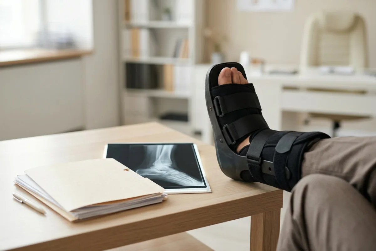
The hindfoot often needs more detail. Because of the complex anatomy of the calcaneus, a CT scan is often ordered after a fracture has been diagnosed on X-ray, the AAOS explains. For the talus, the AAOS says a CT scan may be ordered when the X-rays leave questions, since it helps the doctor see the fracture lines more clearly.
Subtle midfoot injuries can hide on plain films. After a low-energy injury, the AAOS says, the doctor may ask for an X-ray taken while standing to look for a ligament injury, may X-ray the uninjured foot for comparison, and might order an MRI because subtle Lisfranc injuries can be difficult to diagnose. In a 2024 study in Experimental and Therapeutic Medicine of 31 patients with surgically confirmed subtle Lisfranc injuries, non-weight-bearing X-rays made the correct diagnosis in 48.4% of patients, CT in 87.1%, and MRI in 96.8%. The authors concluded that non-weight-bearing X-rays had poor diagnostic accuracy for these injuries.
The page on radiology imaging after a crash compares these tests in more depth, and the page on reading an MRI report walks through common report terms.
Casts, Boots, and Surgical Repair
Treatment for a calcaneus fracture aims to restore the normal anatomy of the heel, and in most cases surgery is the only way to re-create it, according to the AAOS. A fracture whose pieces have not been displaced may be held in a cast, splint, or brace, and a cast may have to be worn for 6 to 8 weeks or possibly longer, with no weight on the foot until the bone is completely healed, the AAOS says. For displaced fractures, the same page describes screws placed through small incisions and open reduction with plates and screws, and notes that surgeons may wait for swelling to go down when the skin is not broken.
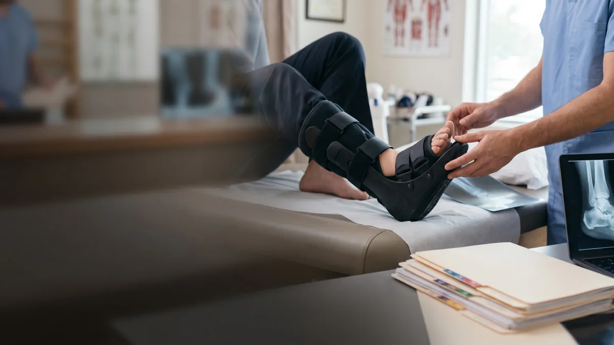
For a Lisfranc injury with no fractures or dislocations and ligaments that are not completely torn, the AAOS describes 6 to 8 weeks in a non-weight-bearing cast or boot. Surgery is recommended for displaced fractures or unstable joints, according to the AAOS, through open reduction and internal fixation with plates or screws, some or all of which may need to be removed 3 to 5 months after surgery, or through a midfoot fusion that lets the injured bones heal together.
Ankle treatment follows the fracture pattern. A nondisplaced medial malleolus fracture can be treated with a short leg cast or walking boot, and a nondisplaced lateral malleolus fracture often can be, according to the AAOS, while most bimalleolar fractures need surgery, usually with a plate and screws or a nail. For a displaced talus fracture, the AAOS says surgery to set and stabilize the broken pieces results in the best outcome and reduces the risk of future complications.
Recovery Ranges by Injury Type
Most ankle fractures take at least 6 weeks for early healing and 10 to 12 weeks for complete healing, according to the AAOS, though damaged ligaments and tendons may take longer. Most people return to normal activities within 3 to 4 months after the injury, the AAOS says. A person whose right ankle is affected can probably resume driving at 9 to 12 weeks, the AAOS adds, and complete recovery from some ankle fractures may take up to 2 years.
For heel fractures, some patients can begin weight-bearing activities a few weeks after injury or surgery and some can begin partial weight-bearing at 6 to 10 weeks, but most need to wait 3 months before putting weight on the heel, according to the AAOS. A minor injury may allow a return to normal activities 3 to 4 months after surgery, the AAOS says, while a severe fracture may take 1 to 2 years before recovery is complete.
After talus fracture surgery, the foot stays in a splint or cast for 2 to 8 weeks, and the AAOS says a person will not be able to put all of their weight on the foot for up to 2 to 3 months. For a Lisfranc injury treated with surgery, weight-bearing in the boot starts after 6 to 12 weeks if the X-rays look appropriate, and resuming all activities may take 6 months to 1 year in some patients, according to the AAOS.
Arthritis, Necrosis, and Lasting Stiffness
Even with appropriate treatment, calcaneus fractures can be devastating and life-changing, the AAOS says, and many may result in long-term complications such as pain, swelling, loss of motion, and arthritis. Patients rarely regain normal foot and ankle motion after a severe fracture, according to the AAOS, and many patients with labor-intensive jobs are unable to return to their job after a calcaneus fracture.
If the bone has healed in a deformed position or the subtalar joint becomes arthritic, the AAOS says the joint between the talus and the calcaneus may need to be fused so the two bones grow together into one. The fusion is very successful in reducing or eliminating pain but results in limited hindfoot motion, the AAOS says. If the arch is not restored or the Achilles tendon does not heal at its normal distance from the ankle, the same page notes, walking on uneven ground such as grass or hills can become a problem.
Talus fractures carry their own long tail. In a 2009 study in International Orthopaedics that followed 20 patients for an average of 7.5 years after surgery for displaced talar neck and body fractures, four patients (20%) developed osteonecrosis of the talus and seven (35%) had undergone secondary surgery by final follow-up. The injured foot in that study showed a significant loss of range of motion compared with the healthy foot, and the AAOS states that a significant percentage of talus fractures result in some degree of posttraumatic arthritis.
After an ankle fracture, many patients have stiffness and swelling for many months even after the fracture has healed, according to the AAOS, which also lists arthritis that can develop years after the injury among the complications of ankle fracture surgery.
Records From Diagnosis to Recovery
For a calcaneus fracture, the AAOS describes doctors checking the pulse at key points of the foot, testing whether the toes move and the bottom of the foot has feeling, and examining the other leg, the pelvis, and the spine for more injuries. The same page stresses telling the doctor as much detail as possible about how the injury happened.
For ankle fractures, the AAOS says the doctor will most likely monitor bone healing with repeated X-rays, typically at 2 weeks, 6 weeks, and 12 weeks. The AAOS warns that putting weight on a healing heel too soon may move the bone pieces out of place or cause surgical screws to loosen or break.
Copies of the emergency X-rays, any later CT or MRI, the operative report, and each written weight-bearing restriction show how a foot or ankle injury changed over time. Kept together with their dates, those records let each new provider see the full course from the first exam forward.
Foot or Ankle Still Hurting After the Crash?
A heel, talus, or midfoot fracture can take many months to settle, and an insurer may treat the first emergency room visit as the whole story. Sam Aguiar Injury Lawyers gathers the X-rays, CT and MRI reports, operative notes, and therapy records so the full course of your foot or ankle injury is on paper. Under our Bigger Share Guarantee®, you always walk away with more than the firm after bills, liens, and case costs. Start with a free case review.