

Car Accident Pelvic Fractures

Key Takeaways
- Pelvic fractures make up only about 3% of all fractures in adults, and most are caused by trauma, typically a high-energy event such as a motor vehicle collision, according to the American Academy of Orthopaedic Surgeons.
- Road traffic accidents typically cause anterior-posterior compression, or open book, fractures, which can lead to pelvic bleeding and hypovolemic shock, a 2022 review in Diagnostics explains.
- Pelvic fractures usually take 8 to 12 weeks to fully heal, and more severe fractures could take longer, especially with other injuries or complications, according to Cleveland Clinic.
Bones of the Pelvic Ring
The pelvis is the ring of bones at the base of the spine, according to the American Academy of Orthopaedic Surgeons (AAOS). It is built from the sacrum, the coccyx or tailbone, and the hip bones. Each hip bone contains three bones, the ilium, ischium, and pubis, which are separate during childhood and fuse together with age. Where those three meet they form the acetabulum, the hollow cup that serves as the socket of the hip joint.
Major nerves, blood vessels, and parts of the bowel, bladder, and reproductive organs all pass through the pelvic ring, and the ring protects them, the AAOS explains. It also anchors the muscles of the hip, thigh, and abdomen. Breaks here are uncommon, making up only about 3% of all fractures in adults.
The bones and joints of the ring have no inherent stability on their own, a 2022 review in Diagnostics points out. The pubic symphysis at the front provides only 15% of pelvic stability, and stability depends mainly, in a ratio of 8:2, on the ligaments of the posterior tension band at the back of the pelvis.
Because the pelvis is shaped like a ring, a break at one point tends to come with damage at a second point. The AAOS says that fracturing one part of the ring often means a fracture or ligament damage somewhere else in it. The Diagnostics review states the rule more firmly, with two exceptions: avulsion fractures in young people and insufficiency fractures in older adults, where even mild trauma can break the ring at a single point.
Crash Forces Behind the Injury
Most pelvic fractures are caused by trauma, typically a high-energy event such as a motor vehicle collision, the AAOS reports. Because the pelvic bones sit near major blood vessels and organs, these fractures may cause extensive bleeding and other injuries that require urgent treatment.
The pelvis is a very stable structure, so most pelvic fractures come from high-impact events such as a car accident or a fall from a significant height, and those events usually cause unstable fractures, Cleveland Clinic notes. Severe pelvic fractures are most common in people aged 15 to 28 years, Cleveland Clinic adds.
In high-energy blunt trauma, pelvic ring fractures are rarely isolated and are often associated with injuries to the brain, lung, spleen, liver, kidney, long bones, and thoracic aorta, according to the Diagnostics review.
United States side impact regulations use two acceleration-based injury criteria, one for the chest and one for the pelvis, with a peak pelvic lateral acceleration threshold of 130 g in regulatory tests, a 2006 review in the Journal of Biomechanics explains. In the sled tests that review describes, pelvic fractures resulted from thinner 75 mm padding, while thicker padding of 100 to 150 mm mitigated trauma.
In frontal crashes, epidemiological studies cited in the Journal of Biomechanics review reported that while frontal airbags protect the head and chest, lower extremity injuries have increased, and the pattern of hip injuries has changed to include fractures of the pelvis and acetabulum without femur or knee injury. Acetabular fractures most commonly occur in high-energy injuries such as car and motorcycle accidents, Cleveland Clinic says. The page on hip injuries after a car accident covers the hip joint itself.
Early Signs of a Broken Pelvis
A fractured pelvis is almost always painful, and moving the hip or trying to walk makes the pain worse, the AAOS says. Patients often hold the hip or knee bent in one position to keep the pain from building, and some have swelling or bruising in the hip area.
Pain in the groin, hip, or lower back, pain that grows more intense when walking or moving the legs, numbness or tingling in the groin or legs, abdominal pain, difficulty urinating, and difficulty walking or standing are the signs Cleveland Clinic lists, noting that symptoms depend on how mild or severe the fracture is.
A break through the hip socket causes severe hip pain that is sometimes worsened with movement, according to Cleveland Clinic. When nerves are damaged, a person may feel numbness or tingling down the leg, along with weakness in that leg.
People with high-energy fractures may also have injuries to the head, chest, abdomen, or legs, the AAOS warns. If those injuries cause significant blood loss, the result can be shock, a life-threatening condition that can lead to organ failure.
Pelvic Fracture Warning Signs
- Pain in the groin, hip, or lower back
- Pain that worsens with moving the hip or trying to walk
- Difficulty standing or walking
- Swelling or bruising in the hip area
- Numbness or tingling in the groin or legs
- Pain in the abdomen
- Difficulty urinating
- Weakness in one leg
Stable and Unstable Ring Patterns
In a stable fracture there is often only one break in the pelvic ring, and the broken ends of the bones line up adequately, according to the AAOS. Low-energy fractures are often stable. Iliac wing fractures, incomplete sacrum fractures, and fractures of the superior and inferior pubic rami are the stable patterns the AAOS names.
An unstable fracture usually has two or more breaks in the ring, and the ends of the broken bones do not line up correctly, which is called displacement. This type is more likely to occur in a high-energy event, the AAOS notes, and its unstable patterns are anterior-posterior compression, lateral compression, and vertical shear.
Any of these can also be open, with bone fragments sticking out through the skin. Open pelvic fractures are serious injuries with a high mortality rate, because once the skin is broken, infection can set in both the wound and the bone, by the AAOS account.
The Young and Burgess classification combines the mechanism of the fracture with its stability, the Diagnostics review explains, and sorts injuries by the direction of force: lateral compression in 50% to 70% of cases, anterior-posterior compression in 20% to 30%, vertical shear in 14%, and a combined mechanism. An older system, proposed by Tile in 1988, gave great importance to stability.
Why Kentucky Hires
Sam Aguiar Injury Lawyers
Lateral Compression Fractures
Lateral compression is the most common cause of pelvic ring fracture and is often seen in pedestrians hit by a car, according to the Diagnostics review. The force rotates half of the pelvis inward and reduces the volume of the pelvis. The AAOS describes the same pattern as one in which the pelvis is pushed inward.
These injuries typically cause bladder rupture and damage to the sacral nerves, while pelvic hemorrhage is less common, the review reports.
Severity climbs in three grades. Grade 1 combines a compression fracture of the sacrum on the side of impact with a transverse fracture of the pubic rami, and the Diagnostics review describes these as stable injuries managed without surgery. Grade 2 adds an iliac wing fracture and widening at the back of the sacroiliac joint to the grade 1 injuries, which leaves the ring rotationally unstable and calls for stable internal fixation. Grade 3, sometimes called a windswept pelvis, pairs the inward force on one side with an outward rotation of the other half of the pelvis, and the resulting multidirectional instability also requires internal fixation.
Lateral compression dominated the 3,314 pelvic ring injuries reviewed in a 2022 study in the Journal of Orthopaedic Trauma from a Level I trauma center: 2,025 were grade 1, 347 were grade 2, and 218 were grade 3.
Open Book Anterior-Posterior Injuries
In an anterior-posterior compression injury, the force usually lands on the pubic symphysis at the front of the pelvis and rotates one or both halves outward, increasing the volume of the pelvis, the Diagnostics review explains. Road traffic accidents typically cause this kind of trauma, and the injuries are called open book fractures.
Pelvic bleeding and hypovolemic shock are possible complications, so open book fractures need quick treatment with pelvic wrapping to reduce the pelvic volume, according to the review. Widening of the pubic symphysis is the defining sign, with a gap of more than 1 cm. A gap wider than 2.5 cm is typically associated with damage to the ligaments of the posterior tension band, which makes the pelvis unstable, and symphysis widening is sometimes associated with bladder and urethral injuries.
The Diagnostics review grades these injuries by that gap. Grade 1 has a symphysis gap under 2.5 cm and is stable, managed without surgery. Grade 2 has a gap over 2.5 cm plus widening at the front of the sacroiliac joint from torn anterior sacroiliac, sacrospinous, and sacrotuberous ligaments, leaving rotational instability that requires stable internal fixation. Grade 3 adds widening at the back of the sacroiliac joint as well, with instability in more than one direction.
In the Journal of Orthopaedic Trauma cohort, 59 injuries were anterior-posterior compression grade 1, 426 were grade 2, and 145 were grade 3.
Vertical Shear and Combined Mechanisms
A force acting in the head-to-foot direction, typically a fall from a height, can destroy the back of the pelvis and shift one half of it upward, the Diagnostics review explains. The AAOS describes a vertical shear fracture the same way, as one half of the pelvis shifting upward. On imaging, the Diagnostics review notes a telltale sign: a pulled-off transverse process of the fifth lumbar vertebra on the side of impact, which marks a torn iliolumbar ligament.
Vertical shear leaves the ring both rotationally and vertically unstable and is associated with vascular, genitourinary, and neurological complications, according to the review. Combined mechanism injuries are rare and occur especially in very high-energy trauma. Lateral compression with vertical shear is the most common combination, and these injuries are unstable and require surgical fixation.
Sacral fractures are part of the pelvic ring and are rarely isolated, the Diagnostics review notes. Under the Denis classification the review describes, a zone I fracture lies in the sacral wing outside the nerve openings and impinges on the L5 or S1 nerve root in 6% of cases, while a zone III fracture involves the central canal and carries neurological injury in 56% of cases, with bowel and genitourinary dysfunction.
Both groups were small in the Journal of Orthopaedic Trauma cohort: 30 vertical shear injuries and 57 combined mechanism injuries out of 3,314.
Pelvic Contusion and Fracture Compared
Contusion is the medical name for a bruise, Cleveland Clinic explains, and a bone contusion is blood trapped under the surface of a bone after an injury. If something hits a bone with enough force, it can bleed without being broken. Bone bruises can affect any bone in the body, and car accidents are among the common causes Cleveland Clinic lists.
Bone pain, tenderness, swelling, and skin bruising or discoloration are the most common symptoms. A bone bruise usually feels like a dull, throbbing ache coming from deeper inside the body, and depending on which bone is bruised, the pain might get worse when a person moves, stands, or sits in certain positions, according to Cleveland Clinic.
X-rays cannot show bone bruises, Cleveland Clinic notes, though a provider might use one to check for a fracture, and MRI, ultrasound, or CT can picture the bone and the area around it. Most bone bruises last a few weeks, while more severe ones can take months or longer to heal. Untreated, a bone contusion can weaken the bone enough to cause a fracture.
The table below sets a bone contusion beside stable and unstable pelvic fractures, drawing on Cleveland Clinic’s bone bruise and pelvic fracture pages and the AAOS page cited above.
| Feature | Bone Contusion | Stable Pelvic Fracture | Unstable Pelvic Fracture |
|---|---|---|---|
| What happens | Blood trapped under the bone surface without a break | Often one break in the ring; the ends line up | Usually two or more breaks; the ends are displaced |
| Typical cause | Falls, sports injuries, car accidents | Often a low-energy event, such as a minor fall in someone with weak bones | More likely a high-energy event such as a car crash |
| Imaging | Not visible on x-ray; MRI, ultrasound, or CT | X-rays from several angles; CT commonly ordered | X-rays and CT to define the pattern and other injuries |
| Common treatment | Rest, ice, compression, elevation; sometimes a splint or brace | Walking aids, pain medication, a blood thinner | External fixation, traction, or plates and screws |
| Healing range | Most last a few weeks; severe ones take months or longer | Usually 8 to 12 weeks | Can take longer, especially with other injuries |
X-Rays, CT Scans, and MRI
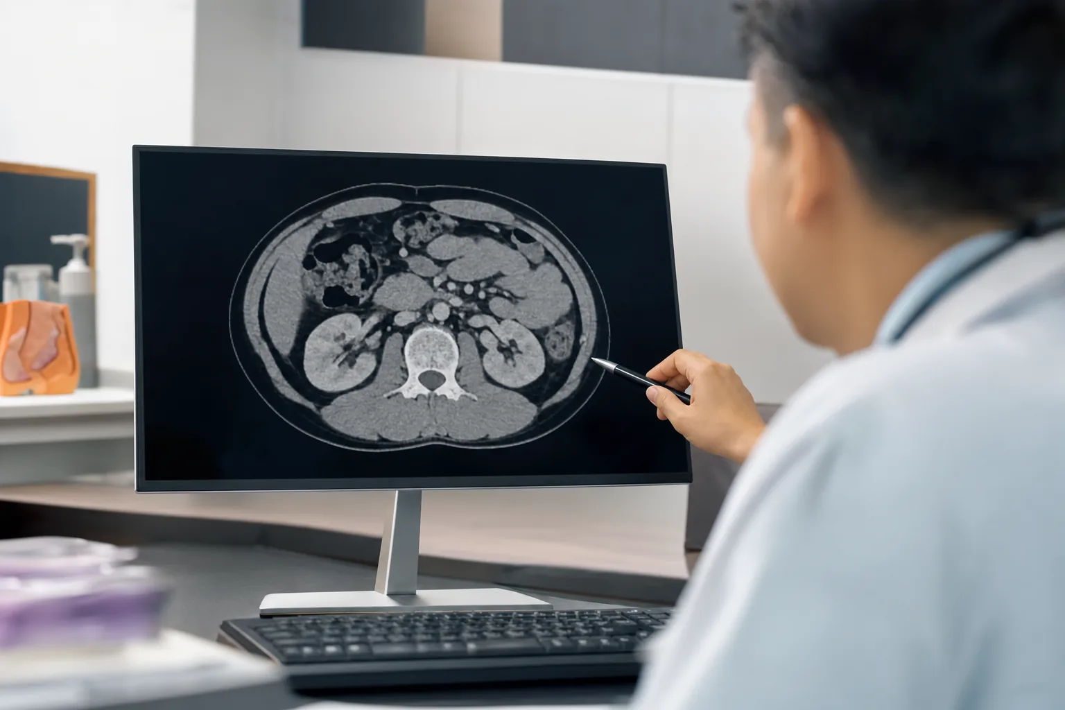
Every pelvic fracture requires x-rays, usually from several angles, to show how far the bones have moved out of place, the AAOS says. Because the injury is complex, a CT scan is commonly ordered to define the pattern and extent of the fracture, look for other injuries, and plan any surgery. In rare cases, an MRI may be ordered to check for fractures that x-ray and CT cannot show.
CT is the gold standard for high-energy blunt trauma when the patient has no hemodynamic abnormalities, according to the Diagnostics review. With the pelvis involved, CT can quickly and accurately identify ring fractures, active bleeding, and bladder rupture. For a patient whose circulation is unstable, a single front-to-back x-ray of the pelvis can still show widening of the pubic symphysis, fractures of the pubic rami, and a pulled-off L5 transverse process.
The extended focused ultrasound exam for trauma, called e-FAST, has low sensitivity for bleeding behind the abdominal lining, but it can point to possible symphysis widening, the review notes. CT run in several phases separates venous from arterial bleeding, which matters because each is managed differently. CT cystography is the most accepted test for bladder injury, and MRI is the best technique for evaluating the lumbar and sacral nerve roots.
The AAOS describes doctors examining the pelvis, hips, and legs, checking for nerve injury by testing whether the patient can move the ankles and toes and feel sensation on the bottoms of the feet, and then examining the rest of the body for other injuries.
Emergency Bleeding Control
Bleeding inside the pelvis is an early complication that leads to hypovolemic shock and early death in 5% to 18% of severe trauma, according to the Diagnostics review. In 80% to 90% of cases the bleeding is venous, from torn veins around the bladder, the presacral venous plexus, or the broken bone itself. In 10% to 20% it comes from branches of the external or internal iliac arteries.
Venous bleeding is often slow and self-limited and can be treated with blood transfusion, pelvic packing, or a pelvic binder, the review explains. Arterial bleeding requires angiographic embolization, or pelvic packing for an unstable patient when angiography is not available.
Patients with high-energy fractures will almost always go to an emergency room first, and their care requires a team of specialists, per the AAOS. In some cases doctors must address airway, breathing, and circulation before treating the fracture. For patients who cannot tolerate a lengthy procedure, an external fixator may serve as a temporary treatment until another procedure can be performed.
At one Level I trauma center, mortality after pelvic ring injury fell from 9.1% in 1999 through 2006 to 4.9% in 2007 through 2018, the Journal of Orthopaedic Trauma study found. The authors note that the improvement coincided with changes in trauma resuscitation and adjuvant treatments for severe hemorrhagic shock, and that mortality for these injuries in the presence of hemorrhagic shock ranges from 40% to 60%.
Surgical Fixation of the Ring
Treatment depends on the specific fracture pattern, how far the bones are displaced, and the patient’s overall condition and other injuries, according to the AAOS. For stable fractures with little or no displacement, nonsurgical care may include crutches or a walker for up to 3 months, pain medication, and a blood thinner to reduce the risk of clots in the veins of the legs and pelvis.
In external fixation, the surgeon inserts metal pins or screws into the bone through small incisions, and the pins stick out on both sides of the pelvis, where they attach to carbon fiber bars outside the skin, the AAOS explains. The frame holds the broken bones in position. Some patients keep it until healing is complete, while others have it as a temporary step.
Skeletal traction uses a pulley system of weights and counterweights, with pins placed in the thighbone or shinbone, to pull the fragments into as normal a position as possible. It is often used right after the injury and removed after surgery. In open reduction and internal fixation, displaced fragments are repositioned and held with screws or metal plates attached to the outer surface of the bone, the AAOS says.
Most acetabular fractures need surgery, according to Cleveland Clinic, either open reduction and internal fixation or, when the socket is weak or too damaged to repair, a total hip replacement. Surgical time varies with the severity of the fracture and may range from two to six hours. For severe pelvic fractures from high-impact accidents, Cleveland Clinic notes that the success of treating the fracture often depends on the success of treating the related injuries.
Recovery Timelines and Walking Aids
Pelvic fractures usually take 8 to 12 weeks to fully heal, Cleveland Clinic reports, and more severe fractures could take longer, especially with other injuries or medical complications from the same event.
After surgery, most patients begin walking with weightbearing restrictions, along with foot and leg exercises, as soon as possible, the AAOS explains. Crutches or a walker may be needed for 6 to 8 weeks or longer, depending on the severity of the injury and how it is healing. Full weightbearing is usually allowed by 3 months or when the bones are fully healed, and a cane or walking aid may be needed for longer.
Stable pelvic fractures tend to heal well, the AAOS says. A person may walk with a limp for several months if the muscles around the pelvis were damaged, and those muscles may take up to 1 year to become strong again. In the Journal of Orthopaedic Trauma cohort, hospital length of stay was 10.6 and 10.7 days in the two study periods.
A hip socket fracture has its own timeline. Partial weight on the leg may be possible after six to eight weeks, while full weight waits a few months, according to Cleveland Clinic. More physical activity has to wait six to 12 months, and many people do not return to the level of activity they had before.
Bladder, Nerve, and Lasting Complications
The bladder and urethra are injured in 3.4% and 1% of pelvic trauma cases, the Diagnostics review reports, and these injuries are more common when the pubic symphysis has widened or broken pubic fragments are displaced. Rupture outside the abdominal lining is the most common bladder injury, accounting for 80% to 90% of cases, and occurs in association with anterior ring fractures; it is treated without surgery, while rupture into the abdominal cavity is rare and needs surgery. Urethral injuries are more common in men. The page on car accident internal organ injuries covers other organs a crash can damage.
Nerve injury is a potential complication, particularly when the sacrum is involved, and it is often overlooked in early trauma care, according to the review. Fractures through the spinal canal or the nerve openings can be associated with nerve root, plexus, or cauda equina damage, and the presentation varies: motor weakness in the foot, loss of sensation, bladder incontinence, fecal incontinence, and sexual dysfunction. The review also notes that testicular trauma can accompany pelvic fractures, especially in motor vehicle crashes and falls from a height.
Surgery carries its own risks. The AAOS lists wound healing problems including infection, damage to nerves or blood vessels, blood clots, pulmonary embolism, nonunion, malunion, and a difference in leg length if the ring heals in an abnormal position on one side.
Pain, reduced mobility, incontinence, and sexual dysfunction may follow from the nerve and organ damage tied to a pelvic fracture, the AAOS warns. After a hip socket fracture, Cleveland Clinic names posttraumatic arthritis, avascular necrosis, sciatic nerve injury that can cause foot drop, and heterotopic ossification, bone growing in the soft tissue around the socket, among the possible complications.
Records That Track the Healing
The first exam on record, including the nerve checks described above, sets the baseline. The fracture grade recorded early, such as lateral compression grade 2 or anterior-posterior compression grade 3, links the injury to the stability findings and complications the Diagnostics review associates with each type.
Later entries follow the bone and the nerves. Plain x-rays, including angled inlet and outlet views, are also important in the follow-up of pelvic fractures, according to the review, and Cleveland Clinic advises contacting a provider about new or concerning symptoms after a pelvic fracture has been diagnosed. Operative reports, therapy notes, and urology or nerve testing fill in the months between. The page on car accident femur fractures covers breaks lower in the leg.
A dated log of when a binder, fixator, wheelchair, walker, or cane was used, and when weight on each leg was first allowed, gives later providers a timeline to check against. Kept with the discharge summary and each set of films, that log puts the whole course of a pelvic injury in one place.
Pelvis Still Hurting Months After the Crash?
A broken pelvis can mean a binder in the trauma bay, a fixator or plates, weeks in a wheelchair or on a walker, and bladder or nerve problems that surface later, and an insurer may judge the whole injury by the discharge papers. Sam Aguiar Injury Lawyers gathers the trauma records, the CT reports, the operative notes, and the therapy and urology records so each stage of your recovery is on paper. Under our Bigger Share Guarantee®, you always walk away with more than the firm after bills, liens, and case costs. Start with a free case review.