

Car Accident Cervical Spine Injuries

Key Takeaways
- Pain from a neck sprain may not appear until hours after an accident, or even the next day.
- Plain x-rays missed 55.5% of clinically significant cervical fractures, which is why CT is usually the first scan after serious neck trauma.
- Most neck sprain symptoms go away in 4 to 6 weeks, yet persistent symptoms remain in 25 to 40% of whiplash patients after 1 year.
Seven Bones That Form the Neck
The cervical spine is the neck section of the spine. It is seven stacked vertebrae, numbered C1 to C7, that support the weight of the head, surround and protect the spinal cord, and allow a wide range of head motion.
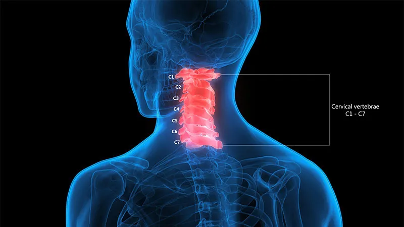
The top two bones are shaped differently from the rest. C1, the atlas, is a ring of bone that holds the head upright, and C2, the axis, lets the atlas pivot for the side-to-side “no” turn of the head. Six disks sit between the seven vertebrae as cushions, and eight pairs of spinal nerves, labeled C1 through C8, exit through small openings to supply movement and feeling to the neck, shoulders, arms, and hands.
Ligaments hold the column together. The anterior longitudinal ligament runs down the front of the vertebrae and stretches to resist backward neck motion, while the posterior longitudinal ligament runs down the back and resists forward motion.
Crash Forces and the S-Shaped Curve
A neck sprain or strain can happen when a sudden movement, such as a motor vehicle accident, bends or twists the neck to an extreme position. One example is whiplash, the neck whipping rapidly back and forth, commonly seen after rear-end accidents.
Whiplash is bony or soft tissue injury from rear-end or side impact, predominantly in motor vehicle accidents, through an acceleration-deceleration transfer of energy to the neck. Recent research points to hyperextension of the lower cervical vertebrae while the upper vertebrae flex, which bends the neck into an S shape at the moment of impact.
More energy can do more than the soft tissue stretch of whiplash. Motor vehicle accidents are among the usual causes of cervical fractures, and they are the leading cause of spinal cord injuries for people younger than 65.
Symptoms That Surface Hours Later
Neck pain does not always start at the scene. Pain from a neck sprain may not appear until hours after the injury, or even the next day, and pain that peaks a day or so after the injury is among the symptoms.
Some neck pain is a warning sign that calls for immediate medical attention: pain that is constant and does not ease over time, severe pain that over-the-counter medication does not relieve, pain that shoots down the arms and legs, and pain with headache and numbness, tingling, or weakness in the arms and legs. For a possible spinal cord injury, the emergency symptoms are below.
Spinal Cord Emergency Signs
- Serious back pain or pressure in the neck, head, or back
- Weakness, loss of coordination, or loss of control in any part of the body
- Numbness, tingling, or loss of feeling in the hands, fingers, feet, or toes
- Loss of bladder or bowel control
- Trouble with balance and walking
- Trouble breathing after the injury
- A twisted neck or back
Sprains and Strains of Soft Tissue
The seven neck vertebrae are tied to each other by ligaments and muscles, which are like thick rubber bands. A sprain is an injury to those ligaments. A strain is an injury to the muscles. Both can follow an accident that bends or rotates the neck in an abnormal way.
Diagnosis starts with the exam. The doctor asks how the injury happened, tests the range of motion of the neck, and checks for point tenderness. A sprain or strain cannot be seen on an x-ray because it involves soft tissue, but an x-ray may be ordered to rule out a fracture, a dislocation, or arthritis, and a CT or MRI may be added in certain cases.
Why Kentucky Hires
Sam Aguiar Injury Lawyers
Pinched Nerves and Herniated Disks
Cervical radiculopathy, usually called a pinched nerve, is compression or irritation of a nerve in the neck where it branches away from the spinal cord. The pain usually starts at the neck and travels down the arm in the area that nerve serves. Patients often describe it as burning or sharp, with tingling in the fingers or hand, weakness in the arm, shoulder, or hand, and numbness.
Other problems can produce the same pattern: some shoulder conditions, heart disease, and nerve entrapment at the elbow or wrist, including carpal tunnel syndrome, can mimic a pinched nerve in the neck.
Fractures of the Cervical Vertebrae
A break in one of the cervical vertebrae is commonly called a broken neck. These fractures usually result from high-energy trauma, such as motor vehicle accidents or falls, and in older people even a ground-level fall can cause one.
A broken neck does not always announce itself with severe pain. Conscious patients with an acute neck injury may or may not have severe neck pain. Some have pain spreading to the shoulders or arms from a vertebra pressing on a nerve, along with bruising and swelling at the back of the neck. Emergency crews treat any unconscious person as if the neck is injured, and the neck is immobilized until x-rays are taken and reviewed by a physician.
Fracture-dislocations of the spine come mostly from vehicle accidents or falls from heights.
Spinal Cord Damage at Neck Level
The spinal cord runs through the center of the cervical vertebrae, so a neck injury can reach it. An injury to the upper, cervical spinal cord results in weakness or paralysis of the arms and legs, and an injury high in the neck can cause severe breathing problems from paralysis of the breathing muscles.
The National Institute of Neurological Disorders and Stroke separates complete from incomplete injuries. With an incomplete injury, the cord can still send some messages, so some feeling, function, and muscle control remain below the injury. With a complete injury, there is no nerve communication below the injury site.
Injuries in the cervical spine lead to more disability than injuries in the thoracic or lumbar spine. A person who recovers some movement or feeling within 1 week usually has a good chance of recovering more function, though that may take 6 months or more, and losses that remain after 6 months are more likely to be permanent.
Three Injury Patterns Side by Side
The table sets the three injury patterns side by side: sprains and strains, pinched nerves, and fractures.
| Feature | Sprain or Strain | Pinched Nerve | Cervical Fracture |
|---|---|---|---|
| Injured tissue | Neck ligaments or muscles | A nerve root where it branches from the spinal cord | One of the seven cervical vertebrae |
| Common signs | Neck pain that worsens with movement, stiffness, headache in the back of the head | Burning or sharp pain down the arm, tingling, weakness, numbness | Neck pain that may or may not be severe, pain into the shoulders or arms, swelling at the back of the neck |
| Plain x-ray | Cannot show the injury itself | Shows alignment, narrowing of nerve openings, and disk damage | Can miss fractures that CT finds |
| Usual course | Most symptoms gone in 4 to 6 weeks | Most patients get better over time | Minor fractures braced for 8 to 12 weeks |
Decision Rules for Emergency Imaging
Not every neck injury gets a scan. Two decision rules, the Canadian C-spine rule and the National Emergency X-Radiography Utilization Study (NEXUS) criteria, were built to help clinicians decide which patients with neck trauma need imaging. The five NEXUS criteria are no tenderness along the back midline of the neck, no evidence of intoxication, a normal level of alertness, no focal neurological deficit, and no painful distracting injury such as a long-bone fracture.
The shift to CT follows the numbers. Plain radiographs missed 55.5% of clinically significant fractures, with sensitivity as low as 52% compared with 98% for CT, and plain radiographs were fully abandoned for clearing the cervical spine.
CT, MRI, and Nerve Conduction Tests
Each test answers a different question. A CT scan can show the shape and size of the spinal canal, its contents, and the bone around it, while MRI can reveal problems with the spinal cord and the nerves leaving the spinal column, spinal degeneration, and disk herniation.
An MRI of the neck can show whether nerve compression comes from soft tissue damage, such as a bulging or herniated disk, and whether the spinal cord or nerve roots are damaged. Electromyography measures the electrical activity of muscles, and nerve conduction studies done alongside it help determine whether symptoms come from pressure on spinal nerve roots or from a nerve compressed somewhere else, such as the elbow or wrist.
MRI is not routine after a clean CT. Routine MRI is not beneficial for clearing the cervical spine after blunt trauma, but it keeps a role when clinical and imaging findings disagree, such as persistent abnormal neurological findings.
Treatment From Collar to Surgery
Sprains and strains usually heal gradually with time and treatment. Treatment includes a soft collar to support the head, pain relievers, muscle relaxants for spasms, and ice and heat. Massage, traction, exercise, and formal physical therapy may follow as the injury improves, under a doctor’s supervision.
For a pinched nerve, initial treatment is nonsurgical: a soft collar worn for a short period, physical therapy, anti-inflammatory medication, a short course of oral corticosteroids, and steroid injections near the nerve, which may lessen swelling long enough for the nerve to recover. Surgery is an option when nonsurgical care has not relieved symptoms after a period of time.
Fracture care depends on which vertebra broke and how. A minor fracture can be treated with a cervical brace worn for 8 to 12 weeks, while a larger or more complex fracture may need traction, surgery, 2 to 3 months in a rigid brace, a halo vest, or a combination. Surgical options include diskectomy, laminectomy, disk replacement, and fusion, which permanently connects vertebrae so the motion between them is gone.
Recovery Ranges by Injury Type
Most symptoms of a neck sprain or strain go away in 4 to 6 weeks, though severe injuries may take longer. For a pinched nerve, most patients get better over time. For some the pain fades in days or weeks, for others it takes longer, and it is common for the symptoms to return at some point.
Some whiplash patients do not recover on that schedule. Persistent symptoms remain in 25 to 40% of patients after 1 year. Older age, female gender, and higher initial pain intensity are tied to a slower course.
Age-Related Wear on Neck Scans
Neck scans often show changes that were there before the accident. Aging disks lose height and water content and grow stiffer, and the body responds by forming bone spurs, which can narrow the openings where nerve roots exit. These changes are normal and occur in everyone.
Nearly half of all people middle-aged and older have worn disks and pinched nerves that cause no painful symptoms, and it is not known why some people develop symptoms and others do not.
Narrowing can also leave the cord less room. Most low-impact spinal cord injuries occur in older people with spinal stenosis, and most of them were not aware they had it.
Records That Track the Injury Over Time
Breaks in care leave treatment gaps in the medical history of a neck injury.
Specific details make each record more useful: where the pain traveled, which movements made it worse, any numbness or weakness in the arms, and which work or household tasks became hard.
Emergency notes, imaging reports, and therapy records together show how a neck injury began and how it changed over the following weeks. Keeping copies of each, along with the police report from the accident, puts that history in one place.
Neck Still Hurting Weeks After the Crash?
A neck injury can make it hard to check a blind spot, lift a child, or sleep through the night, and an insurer may still point to a clean x-ray or an old disk finding on the MRI. Sam Aguiar Injury Lawyers gathers the records from the first scan onward so the full course of your neck injury is on paper. Under our Bigger Share Guarantee®, you always walk away with more than the firm after bills, liens, and case costs. Start with a free case review.