

Modic Changes on MRI After a Car Accident
Key Takeaways
- Modic changes are signal changes in the vertebral marrow next to a disc. Type 1 refers to increased vascularization and bone marrow edema, type 2 to fatty marrow replacement, and type 3 to subchondral bone sclerosis.
- They are common, and they appear without back pain too: the median reported prevalence was 43 percent in patients with non-specific low back pain or sciatica and 6 percent in non-clinical populations.
- A high-intensity zone is a bright spot in the outer ring of a disc. It represents a deep radial tear of the annulus fibrosus, and later investigators showed it is also present in asymptomatic individuals.
Where the Modic Classification Comes From
Modic and colleagues described the pattern in a 1988 study in Radiology. They reviewed magnetic resonance images of 474 consecutive patients referred for lumbar spine MR imaging, and in all cases there was evidence of associated degenerative disk disease at the level of involvement.
The finding is a signal change in the bone, not a structural break. It sits where the disc meets the vertebra above and below it, in the endplate and the marrow just beneath. In the original study, type 1 changes were identified in 20 patients (4 percent) and type 2 in 77 patients (16 percent).
A lumbar MRI report after an accident can list these words next to a line about the disc. What the words mean, how common they are, and what they can and cannot show is the subject of the sections below.
The Three Modic Types
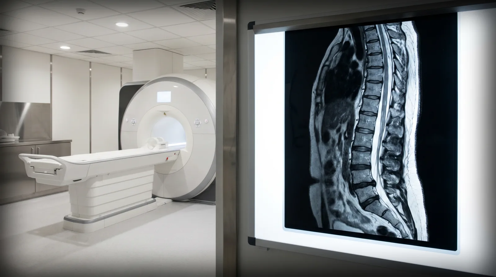
The type describes what the marrow change looks like. Type 1 refers to the presence of increased vascularization and bone marrow edema within the vertebral body, type 2 involves fatty marrow replacement, and type 3 reflects subchondral bone sclerosis.
In the original description, type 1 changes had decreased signal intensity on T1-weighted images and increased signal intensity on T2-weighted images, and type 2 changes had increased signal intensity on T1-weighted images and isointense or slightly increased signal intensity on T2-weighted images.
| Type | What it reflects | Signal in the original study |
|---|---|---|
| Type 1 | Bone marrow edema and increased vascularization | Lower on T1 images, higher on T2 images |
| Type 2 | Fatty marrow replacement | Higher on T1 images, equal or slightly higher on T2 images |
| Type 3 | Subchondral bone sclerosis | Not part of the 1988 description |
Under the microscope, type 1 changes showed disruption and fissuring of the end plates and vascularized fibrous tissue, while type 2 changes showed yellow marrow replacement.
The types are not fixed labels. Research has supported the notion that Modic changes possess a transitional nature, and in the original study type 1 changes in five of six patients converted to a type 2 pattern in 14 months to 3 years, while type 2 changes in ten patients remained stable over a 2 to 3 year period. Type 1 changes have been strongly associated with inflammation and severe low back pain, while types 2 and 3 tend to be more stable and demonstrate less refractory pain.
High-Intensity Zone on T2
A high-intensity zone is a separate finding in a different place. It is a bright focus on a T2-weighted image within the annulus fibrosus, the outer ring of the disc. Aprill and Bogduk described it in 1992: it occurred in 28 percent of 500 patients undergoing MRI for back pain, and its presence correlated significantly with Grade 4 annular disruption and with reproduction of the patient’s pain.
In the same 1992 study, sensitivity was modest but specificity was high, with a positive predictive value of 86 percent for a severely disrupted, symptomatic disc. Later work complicated that: correlation with CT discography showed that the high-intensity zone represents a deep radial tear of the annulus fibrosus, which may be a cause of chronic low back pain, but later investigators demonstrated that it is also present in asymptomatic individuals.
A meta-analysis of adults 50 and younger did not find it tied to back pain: high-intensity zone was among the findings not associated with low back pain, with an odds ratio of 2.10 and a confidence interval from 0.73 to 6.02.
How Common Modic Changes Are
Across 77 articles in a systematic review, the median prevalence of vertebral endplate signal changes was 43 percent in patients with non-specific low back pain or sciatica and 6 percent in non-clinical populations. The prevalence was positively associated with age, and there was a positive association between these changes and low back pain in seven of ten studies, with odds ratios from 2.0 to 19.9.
The caution that matters for a report is that vertebral endplate signal changes may be present in individuals without low back pain. Modic changes are closely related to the normal degenerative process affecting the lumbar spine, with prevalence increasing with age, and their relation to low back pain remains unclear.
A meta-analysis of adults 50 and younger split the picture by type, comparing people with and without back pain.
| Finding | Odds ratio | Associated with back pain |
|---|---|---|
| Modic type 1 changes | 4.01 | Yes |
| Any Modic change | 1.62 | No |
| High-intensity zone | 2.10 | No |
| Disc extrusion | 4.38 | Yes |
| Disc protrusion | 2.65 | Yes |
Modic 1 changes were more prevalent in adults with back pain, while any Modic change and the high-intensity zone were not associated with low back pain. The underlying problem is simple: imaging features of spine degeneration are common in symptomatic and asymptomatic individuals.
Reading the Findings in Context
A scan shows the spine on the day it was taken. It does not carry a date for when a finding began, and because the same findings turn up in people without pain, a line in a report is a description, not a conclusion about an accident. Disc degeneration shows on imaging in 37% of 20-year-olds and 96% of 80-year-olds with no back pain. The type can change over time, as the 1988 follow-up of patients showed, which is why a single study is a snapshot.
What a report line becomes depends on what surrounds it: the symptoms that prompted the scan, the examination, the treatment that followed, and any earlier imaging to compare against. Herniation, protrusion, extrusion, and sequestration are shape words, not treatment orders, and a herniated disc is one where all or part of the disc is forced through a weakened area of its outer ring.
That is also why radiology records carry weight in an injury case, and why pre-existing conditions are handled with the earlier records in hand.
Why Kentucky Hires
Sam Aguiar Injury Lawyers
Pain and Treatment Evidence
Researchers disagree about how much a Modic change matters. Vertebral bone marrow lesions visualized as Modic changes on MRI have a high specificity for discogenic low back pain, and Modic changes are likely to be more than just a coincidental imaging finding in low back pain patients.
Treatment evidence is thin: there is no consensus on the ideal therapy, and non-surgical approaches have some demonstrated efficacy in mostly non-replicated clinical studies in reducing Modic changes in the short term, but with unknown long-term benefits. Strategies under study include rehabilitation, surgical fixation, stabilization, steroid or cement injection, or antibiotics.
Records That Give an MRI Finding Context
The record around the imaging is what gives it meaning, and it is assembled over time. The images in their original form show the sequences and dates, where a report summary does not. Earlier spine imaging, where it exists, allows the comparison that a single study cannot. The ordering clinician’s records show which symptoms prompted the scan, and the treatment before and after shows what changed.
The images on disc or portal rather than only the report, any earlier spine imaging, and a short weekly note on where the back pain sits and what it stops you from doing fill the space between medical visits. Those everyday details fade first.
Back Pain After the Accident and a Confusing MRI Report?
Car accident back pain can come with an MRI report full of terms like Modic changes and high-intensity zone, and an insurer may read those words as a sign of an older condition. You focus on getting better. We handle everything else. With our Bigger Share Guarantee®, you always get more, and you pay $0 out-of-pocket forever. Start with a free case review.