

Knee Injuries After a Car Accident
Key Takeaways
- The American Academy of Orthopaedic Surgeons lists a bent knee hitting the dashboard in a car accident as a common cause of posterior cruciate ligament injury.
- An x-ray will not show a meniscus tear, and the AAOS calls MRI the preferred method for diagnosing acute tears because of its high accuracy.
- In a 2020 study in BMC Musculoskeletal Disorders of 539 adult pedestrians struck by a motor vehicle, 12.4% had ligament injuries in the knee.
Knee Joint Anatomy
The American Academy of Orthopaedic Surgeons (AAOS) calls the knee the largest joint in the body and one of the most easily injured. Three bones meet there: the femur (thighbone), the tibia (shinbone), and the patella (kneecap), which sits in front of the joint as a partial shield.
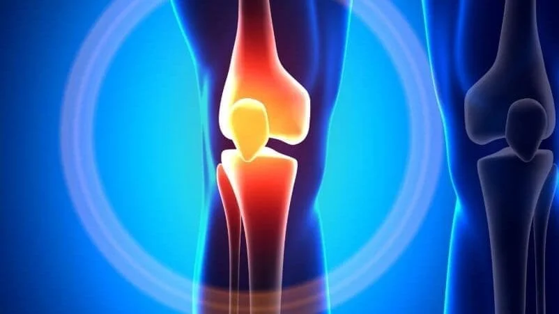
Two wedge-shaped pieces of cartilage, the menisci, sit between the femur and tibia and work as shock absorbers. The AAOS notes that when people talk about torn cartilage in the knee, they usually mean a torn meniscus.
Four main ligaments hold the bones together, according to the AAOS. The medial and lateral collateral ligaments run along the inner and outer sides and control sideways motion. The anterior and posterior cruciate ligaments cross inside the joint in an X and control front and back motion. Tendons finish the picture: the quadriceps tendon attaches the front thigh muscles to the kneecap, and the patellar tendon runs from the kneecap to the tibia.
Dashboard Impact in Frontal Crashes
The AAOS lists a bent knee hitting the dashboard in a car accident as a common cause of posterior cruciate ligament injury, and it describes the injury as one that requires a powerful force. The same blow can break the kneecap: the AAOS page on patellar fractures names a head-on collision that drives the kneecap into the dashboard as one of the most common causes.
Force from the dashboard does not stop at the knee. A 2018 narrative review in African Health Sciences on lower limb injuries in frontal crashes named the knee striking the dashboard as one of the most commonly reported mechanisms, with the impact causing a fracture of the hip socket or a posterior hip dislocation. The authors also described how a driver’s right leg, pressed on the brake, can leave the femur out of position so the knee meets the dashboard.
In a 2020 study in BMC Musculoskeletal Disorders of 539 adult pedestrians struck by a motor vehicle and treated at a level 1 trauma center, 12.4% had ligament injuries in the knee. The authors concluded that clinicians need a high index of suspicion when evaluating these patients.
Cruciate and Collateral Ligament Injuries
The anterior cruciate ligament (ACL) keeps the tibia from sliding forward under the femur and gives the knee rotational stability. The AAOS counts an ACL tear among the most common knee injuries, affecting more than 200,000 people a year in the United States, and lists direct contact or collision among its causes. ACL tears often come with damage to the medial collateral ligament or the menisci.
The posterior cruciate ligament (PCL) is stronger than the ACL and is injured far less often. According to the AAOS, PCL injuries are often subtle and harder to evaluate than other knee ligament injuries, they frequently occur alongside damage to cartilage, other ligaments, and bone, and many are partial tears with the potential to heal on their own.
Sideways force hits the collateral ligaments. The AAOS says a blow to the outside of the knee usually injures the medial collateral ligament, while a blow to the inside that pushes the knee outward can injure the lateral collateral ligament, which tears less often. Doctors grade ligament injuries as sprains: Grade 1 is a mild stretch, Grade 2 is a partial tear that leaves the ligament loose, and Grade 3 is a complete tear with an unstable joint, as the AAOS explains.
Meniscus Cartilage Tears
The AAOS lists meniscus tears among the most common knee injuries and says they can come from acute trauma or from degenerative changes over time. Tears are described by their shape and location, and common patterns include bucket handle, flap, and radial tears.
The early course can mislead. The AAOS says a person may feel a pop, and most people can still walk on the injured knee. Over the next 2 to 3 days, the knee gradually becomes stiffer and more swollen. The most common symptoms are pain, stiffness and swelling, catching or locking, a sense of the knee giving way, and loss of full range of motion.
Age adds a second layer. Worn tissue tears more easily, and the AAOS notes that an awkward twist while getting up from a chair can be enough to tear an aging meniscus.
Early Signs of Internal Damage
Pain and swelling are the most common signs of a knee injury, according to the AAOS. The knee may also catch or lock, and some injuries, an ACL tear among them, cause instability, a feeling that the knee is giving way.
The timing of the swelling carries information. With an ACL tear, the AAOS says the knee swells within 24 hours, and a person may hear a popping noise at the moment of injury. With a PCL injury, the AAOS describes pain with swelling that comes on steadily and quickly, enough to stiffen the knee and cause a limp.
Some signs point to bone or blood flow. The AAOS lists the inability to straighten the knee or hold it straight in a leg raise as a sign of a kneecap fracture. For a break at the top of the tibia, the AAOS says a pale or cool foot may mean blood flow to the foot is impaired, that numbness around the foot raises concern about nerve injury or excessive swelling, and that anyone with these symptoms after an injury should go to the nearest emergency room.
Knee Warning Signs
- A popping noise and the knee giving out at the moment of injury
- Severe pain, or a knee that cannot move
- Swelling at the injury site or a new limp
- Inability to straighten the knee or hold it straight in a leg raise
- A knee that looks out of place
- Pain that gets worse when weight is placed on the leg
- A pale or cool foot below the injured knee
- Numbness or pins and needles around the foot
Why Kentucky Hires
Sam Aguiar Injury Lawyers
Kneecap Fractures From the Dashboard
The kneecap is the bone most commonly broken around the knee, according to the AAOS. The AAOS sorts these fractures into four types: stable fractures, where the pieces stay in contact; displaced fractures, where the ends separate and the joint surface may be disrupted; comminuted fractures, where the bone shatters into many pieces; and open fractures, where bone breaks through the skin and the risk of infection rises.
Bleeding inside the joint is common. The AAOS describes hemarthrosis, blood from the broken bone collecting in the joint space and causing painful swelling, and says a doctor may drain a large collection. X-rays from several angles show the fracture and the alignment, and a CT scan can map multiple fragments before surgery.
Most kneecap fractures need an operation. The AAOS says the pieces move out of place in most patellar fractures, and the strong thigh muscles attached to the top of the kneecap can pull them apart during healing. Transverse fractures are most often fixed with screws, pins and wires, a figure-of-eight tension band, or sutures. A simple, nondisplaced fracture may heal in a cast or splint.
Tibial Plateau and Distal Femur Breaks
The top of the shinbone, where it widens to form the lower half of the knee joint, is called the tibial plateau. The AAOS says young people often break the upper tibia in high-energy injuries such as motor vehicle accidents. The femur is driven into the softer bone of the plateau like a die punch, leaving the surface compressed and sunken, and the skin, muscle, nerves, blood vessels, and ligaments around it may be injured at the same time.
The bottom of the thighbone breaks in a similar setting. The AAOS says distal femur fractures in younger patients usually come from high-energy injuries such as motor vehicle collisions, and that many of these patients also have injuries to the head, chest, abdomen, pelvis, spine, or other limbs. When the bone breaks, the thigh muscles contract and pull the fragments out of line, which makes the fragments hard to line up with a cast.
When surgery is needed, the AAOS describes plates and screws or an intramedullary rod, a temporary external frame when the soft tissue is badly damaged, and bone graft to fill the hole left when a sunken joint surface is lifted back into place.
Dislocation and Multi-Ligament Tears
A dislocated knee is a different order of injury. The AAOS says that in people with normal knee structure, dislocations are most often caused by high-energy trauma such as motor vehicle crashes. In a dislocation, the femur and tibia are forced out of alignment, completely or partially.
The AAOS warns that combined ligament injuries can disrupt the blood supply to the leg, affect the nerves that run the leg muscles, and in severe cases lead to amputation. It says anyone with a suspected multiple ligament injury needs a thorough examination, and that the orthopaedic surgeon may call in a vascular surgeon or microsurgeon.
Surgery tends to come early. For a dislocated knee with several torn ligaments including the PCL, the AAOS says surgery is almost always necessary. The AAOS page on combined ligament injuries adds that more than one operation may be needed, and that outcomes after multiple ligament surgery are not as consistent as after single ligament surgery.
Three Knee Injuries Compared
The table below sets three common crash injuries side by side, using the AAOS descriptions cited on this page. They often overlap: an ACL tear can come with a meniscus tear, and a kneecap fracture can come with damage elsewhere in the leg.
| Feature | Meniscus Tear | ACL Tear | Kneecap Fracture |
|---|---|---|---|
| What is injured | Cartilage cushion between the femur and tibia | Ligament that stops the tibia sliding forward | The patella, the bone at the front of the knee |
| Typical signs | Stiffness and swelling over 2 to 3 days, catching or locking | Pop, giving way, swelling within 24 hours | Front knee pain and swelling, trouble straightening the leg |
| Shows on x-ray | No | No, except a bone fragment pulled loose | Yes |
| Common treatment | Therapy, partial meniscectomy, or repair | Therapy or reconstruction with a tendon graft | Cast or splint if stable, surgery if displaced |
| Usual return | 3 to 6 weeks after trimming, 3 to 6 months after repair | Full sports within 6 to 12 months after surgery | Normal activities within 3 to 6 months |
X-Rays, CT, and MRI Findings
A normal x-ray does not clear the knee. The AAOS says an x-ray will not show a meniscus tear and calls MRI the preferred method for diagnosing acute tears because of its high accuracy. The AAOS says x-rays do not show ligament injuries either, although they can catch an avulsion fracture, a small piece of bone pulled away where a ligament attaches.
Timing affects what the scan shows. For PCL injuries, the AAOS notes that x-rays and MRI can appear normal, especially when the injury happened more than 3 months before the tests. For tibial plateau fractures, the AAOS says CT adds detail about severity, and an MRI may be ordered when the signs point to a fracture but x-rays are negative, because a reaction in the bone marrow can reveal the break.
MedlinePlus says a knee MRI uses strong magnets rather than radiation and most often lasts 30 to 60 minutes. Reasons to order one include a knee that gives way or locks, signs of damage to the meniscus, cartilage, or ligaments, and knee pain that does not improve with treatment. MedlinePlus also lists degeneration and changes that occur with age among the possible causes of abnormal results. The page on reading an MRI report explains common report terms.
Treatment From Bracing to Surgery
Many knee injuries start with simple measures. The AAOS lists a brace or cast to hold the knee still, crutches to keep weight off the leg, physical therapy to restore function and strength, and anti-inflammatory medication. It also says many fractures and injuries around the knee require surgery to fully restore function, with arthroscopic surgery for many ACL tears and open surgery for many fractures.
For the ACL, the AAOS says nonsurgical care is always an option and that reconstruction is the most common surgical treatment. Its page on ACL surgery explains that the torn ligament is generally rebuilt with a tendon graft taken from the patient or from a donor, and that for an acute isolated ACL tear the AAOS recommends 12 weeks of nonsurgical treatment and, if appropriate, surgery within 5 months to prevent more meniscus and cartilage damage.
The meniscus is split by blood supply. The AAOS says the outer third has a rich blood supply, so a tear there may heal on its own or be repaired, while the inner two-thirds cannot heal, so symptomatic tears there that do not respond to conservative care are usually trimmed. The options are a partial meniscectomy, which removes the damaged tissue, or a repair that stitches the torn pieces together.
Recovery Ranges by Injury Type
Recovery depends on the structure and the repair. The AAOS puts rehabilitation after a meniscus repair at about 3 to 6 months and after a partial meniscectomy at roughly 3 to 6 weeks. After ACL reconstruction, its ACL surgery page says most patients return to full sports within 6 to 12 months. For PCL surgery, the AAOS says therapy begins 1 to 4 weeks after the procedure and full recovery typically takes 6 to 12 months.
Fractures run longer. The AAOS says most people return to normal activities within 3 to 6 months after a kneecap fracture, with severe fractures taking longer, and that the knee is often kept straight for 2 to 4 weeks. Full weightbearing after a tibial plateau fracture may take 3 months or more of healing, according to the AAOS, and the AAOS says a distal femur fracture may need 1 year or more of rehabilitation before a return to all everyday activities.
Combined injuries recover slowly, the AAOS notes, and for distal femur fractures the AAOS lists age, bone quality, and the severity of the break among the factors that shape healing.
Post-Traumatic Arthritis and Stiffness
The AAOS defines posttraumatic arthritis as arthritis that develops after an injury to the knee. A broken bone can damage the joint surface and lead to arthritis years later, and meniscus tears and ligament injuries can cause instability and extra wear that, over time, result in arthritis.
Fractures carry specific long-term risks. After a kneecap fracture, the AAOS says severe arthritis develops in a small share of patients and milder arthritis is much more common. Some loss of motion and long-term pain in the front of the knee are also common, and some patients are left with permanent weakness in the thigh muscle. For distal femur fractures that enter the joint, the AAOS says the healed surface may carry a defect that wears down the cartilage, and severe arthritis may eventually be treated with a total knee replacement.
Stiffness is its own problem. The AAOS says some knee stiffness is expected after a distal femur fracture and that bone can heal slowly or not at all, and the AAOS notes that early surgery for multiple torn ligaments can be complicated by scar tissue inside the joint that limits motion.
Medical Records That Track Healing
A knee injury is documented in layers. The first exam compares the injured knee with the healthy one, the AAOS explains, and the AAOS describes checking for tenderness along the joint line and running the McMurray test, which bends, straightens, and rotates the knee to stress a torn meniscus. Each of those findings lands in the chart.
Later records show how the injury behaved. Imaging reports, physical therapy notes, operative reports, and follow-up visits trace whether the knee improved, stalled, or developed stiffness or instability. The page on car accident hip injuries covers the hip and pelvis injuries that can travel up the same leg from a dashboard impact.
Knee symptoms that build over the days after a crash are easier to trace when the emergency note, each imaging report, and every orthopedic visit are kept together. A dated log of swelling, locking, or the knee giving way gives each new provider a record to compare against.
Knee Still Giving Out After the Crash?
A torn meniscus or ligament can hide behind a clean emergency room x-ray, and an insurer may point to that film, or to the word degenerative on a later MRI, to say the crash never touched your knee. Sam Aguiar Injury Lawyers lines up the emergency records, the MRI, and the orthopedic notes so the timeline of your symptoms is on paper. Under our Bigger Share Guarantee®, you always walk away with more than the firm after bills, liens, and case costs. Start with a free case review.