

Car Accident Shoulder Injuries

Key Takeaways
- Plain x-rays do not show the rotator cuff, so x-rays of a shoulder with rotator cuff pain are usually normal.
- Among 664 village residents screened with ultrasound, 22.1% had full-thickness rotator cuff tears and 65.3% of those tears caused no symptoms.
- Nonsurgical treatment relieves pain and improves function in about 80 to 85% of patients with a rotator cuff tear.
Anatomy of the Joint
Three bones meet at the shoulder: the humerus (upper arm bone), the scapula (shoulder blade), and the clavicle (collarbone). Ligaments, tendons, muscles, and a joint capsule hold them together as a platform for the arm. The main joint is the glenohumeral joint, a ball formed by the head of the humerus resting on the golf-tee-shaped glenoid of the shoulder blade.
The socket is shallow for the size of the ball. A rim of cartilage called the labrum surrounds the socket, deepens it by up to 50%, and anchors several stabilizing ligaments along with one of the biceps tendons.
The tendons of four muscles wrapped around the ball form the rotator cuff, which attaches the humerus to the shoulder blade and lifts and rotates the arm. The shoulder is the most mobile joint in the human body, and that mobility makes it an easy joint to dislocate.
Crash Forces on the Shoulder
Motor vehicle collisions are a named cause of several shoulder injuries. A direct blow in a collision can fracture the clavicle or the top of the humerus. The scapula is harder to break because the chest and surrounding muscles protect it, so its fractures usually come from high-energy trauma such as a high-speed crash, and they often occur along with chest injuries.
Falls and motor vehicle collisions are typical causes of a dislocated shoulder, and the most severe dislocations, such as those from vehicle crashes, can also damage nerves or blood vessels.
Traumatic causes of a labral tear include a direct blow to the shoulder and a violent overhead reach, such as an attempt to stop a fall. An acute rotator cuff tear can occur alongside a broken collarbone or a dislocated shoulder. A shoulder belt loads its force across the chest and collarbone in a frontal impact.
Rotator Cuff and Labral Tears
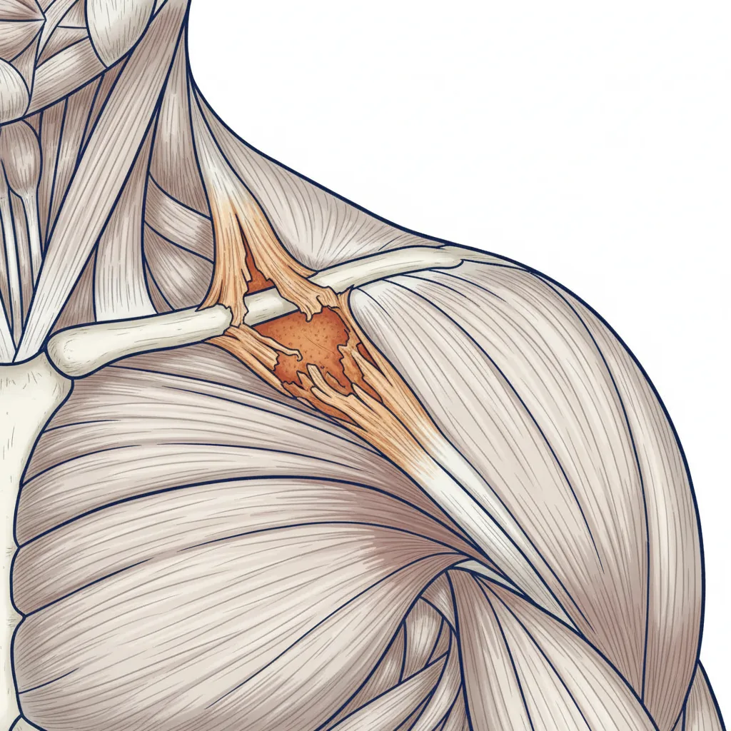
In a rotator cuff tear, one or more tendons pull partly or completely away from the head of the humerus. Most tears involve the supraspinatus tendon. A partial tear leaves the tendon attached but thinner than normal. A full-thickness tear detaches part of it, and in a complete tear there is essentially a hole in the tendon.
A fall on an outstretched arm or a heavy lift with a jerking motion can tear the cuff, while most tears come from slow wear that increases with age. In young adults, most tears are caused by a traumatic injury. The page on car accident rotator cuff injuries covers these tears in more depth.
Labral tears are named for where they sit on the socket rim. A SLAP lesion is a tear on the upper part of the socket that may involve the starting point of the long head of the biceps tendon. A Bankart lesion is a tear at the front and bottom of the socket and usually follows a dislocation in which the ball comes out the front. A tear at the back of the socket is called a posterior labral tear and can result from injury or from wear.
Fractures, Dislocations, and AC Separations
Clavicle fractures make up about 5% of all adult fractures, and are most often caused by a direct blow to the shoulder, including in a motor vehicle collision. Most break in the middle of the bone. The broken ends may stay lined up or move far out of place, which surgeons call a displaced fracture.
A separated shoulder involves the acromioclavicular (AC) joint, where the collarbone meets the highest point of the shoulder blade, and not the main ball-and-socket joint. A Grade I injury is a sprain that looks normal on x-rays. A Grade III injury completely tears both the AC and coracoclavicular ligaments and leaves a larger bump, and Grades IV through VI describe the collarbone pushed backward, upward, or downward, with the downward Grade VI injury described as rare.
Most dislocations push the ball forward out of the socket, which happens when the arm is raised away from the body. When the ball comes out, the socket and the ligaments at the front of the shoulder are often injured, and the trauma can be severe enough to break the glenoid or the upper arm bone as well.
Pain, Weakness, and Loss of Motion
Pain, swelling and bruising, an inability to move the shoulder, a grinding sensation called crepitus, and a visible deformity are the general signs of a shoulder fracture, dislocation, or separation. Each injury adds its own: a bump on top of the shoulder with an AC separation, a bump on the front and an arm rotated outward with a dislocation, and severe swelling with very limited movement after a fracture at the top of the humerus. With a broken collarbone, the shoulder sags downward and forward.
Rotator cuff tears from a sudden event usually cause intense pain, sometimes with a snapping sensation and immediate weakness in the upper arm. The common complaints are pain at night when lying on the injured side, pain when lifting and lowering the arm, and weakness when lifting or rotating it. Some tears are not painful but may still cause arm weakness.
Labral tears are harder to pin down from symptoms alone. They resemble other shoulder injuries and can include deep pain with overhead or reaching activities, catching, locking, popping, or grinding, and a sense that the shoulder is unstable.
Shoulder Red Flags
- A shoulder that looks deformed or out of place
- An arm that cannot be moved or feels like a dead arm
- A hand that is cool to the touch or turns white or purple
- Numbness or weakness in the shoulder or arm
- Broken bone pressing against the skin or breaking through it
- Severe swelling with very limited shoulder movement
Why Kentucky Hires
Sam Aguiar Injury Lawyers
Three Common Injuries Side by Side
Only one of these three is a broken bone, so the first day can look different for each: x-rays show dense structures like bone, which is why a torn tendon or labrum can sit behind a normal film.
| Feature | Rotator Cuff Tear | Labral Tear | Clavicle Fracture |
|---|---|---|---|
| What is injured | Tendon pulled away from the head of the humerus | Cartilage rim around the shoulder socket | The collarbone, most often in its middle portion |
| Traumatic cause | Fall on an outstretched arm, jerking lift, or with a fracture or dislocation | Direct blow, sudden pull, or dislocation | Direct blow to the shoulder, including in a vehicle collision |
| Plain x-ray | Usually normal | Shows bone only; MRI is the best choice for the labrum | Shows the location and severity of the break |
| Usual first treatment | Nonsurgical care, effective in about 80 to 85% | Physical therapy in many cases; SLAP tears typically arthroscopic | Sling, with surgery for badly displaced breaks |
Imaging Beyond the First X-Ray
Most shoulder fractures are diagnosed with x-rays and a physical exam, with CT or MRI added in some cases. For the rotator cuff, x-rays do not show the soft tissues, so x-rays of a shoulder with rotator cuff pain are usually normal. They are taken to rule out other causes of pain, such as arthritis.
Shoulder MRI gives clear views of rotator cuff tears, biceps tendon injuries, and damage to the glenoid labrum, uses no radiation, and can help determine which patients with shoulder injuries require surgery. MRI or ultrasound shows the location and size of a cuff tear, and the accuracy of ultrasound depends on the technician performing the scan.
In direct MR arthrography, a radiologist injects a gadolinium contrast material into the joint so it outlines the cartilage, labrum, ligaments, and bones. Direct arthrography is particularly effective in the shoulder after a dislocation and is often performed only when a standard exam was not enough to assess the problem.
For a dislocation, an MRI is rarely required at the time of the dislocation, and a CT or MRI may not be ordered until the patient sees an orthopaedic surgeon. The page on reading an MRI report explains the terms that appear most often in these reports.
Age-Related Findings on Shoulder MRI
Rotator cuff tears turn up in people who have no shoulder complaints. Ultrasound of both shoulders of 664 residents of one village in Japan found full-thickness tears in 22.1%. The rate was 0% from the 20s through the 40s, 10.7% in the 50s, and 36.6% in the 80s, and 65.3% of the tears caused no symptoms.
A cuff tear did not always cause shoulder symptoms, especially in older people. The screening had limits: the village population was aging, the average participant was 70, and partial-thickness tears were not counted.
Most tears are the result of slow wear that is relatively painless in most cases, and a person with a degenerative tear in one shoulder is more likely to have a tear in the other shoulder even without pain there. MRI can give a better idea of how old or new a tear is because it shows the quality of the rotator cuff muscles.
Conservative Care Without Surgery
Most rotator cuff tears are first treated without an operation. Nonsurgical care relieves pain and improves function in about 80 to 85% of patients. The options include rest and limits on overhead activity, anti-inflammatory medication, physical therapy, and a steroid injection when other measures have not worked, with injections relieving pain in about two-thirds of patients for at least 3 months. The tradeoff is that a tear may grow larger over time.
Most people with an AC separation, professional athletes included, return to normal function without surgery even when a bump remains, and nonsurgical care may be better than surgery for most types. A dislocated shoulder is usually put back in place in the emergency room after sedation and pain medication, then held in a sling.
Operations for Tears and Fractures
Continued pain is the main reason for rotator cuff surgery. Other signs are symptoms lasting 6 to 12 months, a tear larger than 3 cm with good surrounding tissue, significant weakness and loss of function, and a tear caused by a recent, acute injury. The repair most often reattaches the tendon to the head of the humerus.
SLAP tears are typically treated arthroscopically, while Bankart and posterior tears are usually repaired by reattaching the labrum and ligaments with suture anchors. When the injury includes fractures or bone loss, the surgeon may replace the lost bone with bone from elsewhere in the shoulder or from a donor.
For a displaced clavicle fracture, open reduction and internal fixation, which holds the realigned pieces with plates and screws, is the procedure most often used. A fracture at the top of the humerus may be fixed with plates, screws, or pins or treated with a shoulder replacement, and repeated dislocations usually require surgery to repair the torn soft tissues.
Recovery Ranges by Injury Type
Daily life after a shoulder fracture, separation, or dislocation can be greatly affected for several weeks or even months, with most injuries needing a period of immobilization and then rehabilitation. After a less severe injury, function improves quickly once the first 4 to 6 weeks pass, and pain, strength, and range of motion often keep improving for up to 12 months.
A broken collarbone can take several months to heal with or without surgery, and longer in people with diabetes or who use tobacco. Most people return to their regular activities within 3 months of the injury.
After labral repair, a sling is worn for 2 to 6 weeks, with lifting restrictions of no more than 5 pounds for up to 12 weeks, and the shoulder should be fully healed in 4 to 6 months. Most patients regain most of their strength and range of motion.
Stiffness, Instability, and Other Complications
Frozen shoulder, also called adhesive capsulitis, can develop after the shoulder has been immobilized because of surgery, a fracture, or another injury. It moves through freezing, frozen, and thawing stages, and full recovery may take up to 3 years.
A shoulder that has dislocated once is more likely to do it again. People with past dislocations have a high risk of future ones, which can happen even without an injury, and repeated dislocation leads to chronic shoulder instability.
A clavicle fracture that does not heal is called a nonunion; some cause little pain, while others cause pain and arm dysfunction and may need an operation. Risks specific to clavicle surgery include lung injury, numbness below the collarbone, and hardware irritation, and some patients with a plate find that seatbelts irritate the collarbone area. For rotator cuff surgery, permanent stiffness and a repeat tear of the tendon are among the risks.
Medical Records That Track the Injury
For the first shoulder exam, the doctor checks for tenderness, measures range of motion in several directions, tests arm strength, and examines the neck to rule out a pinched nerve. For a dislocation, the doctor needs to know how it happened and whether the shoulder has ever dislocated before.
Imaging reports, the orthopaedic consultation, therapy notes, and any operative report each capture the shoulder at one point in time, and a prior shoulder MRI, if one exists, gives a point of comparison for new findings. The page on treatment gaps explains how breaks in care show up in that record.
A normal first x-ray and a later MRI or orthopaedic finding can sit weeks apart in the chart. Keeping the emergency record, each imaging report, and the therapy notes together shows how the diagnosis developed over that time.
Shoulder Still Hurting Months After the Crash?
A torn rotator cuff or labrum can keep you from lifting, reaching overhead, or sleeping on your side, and an insurer may point to a normal first x-ray or to age-related wear on the MRI to say the crash did little. Sam Aguiar Injury Lawyers lines up the emergency records, the imaging, and the orthopaedic and therapy notes so the course of your shoulder injury is on paper. Under our Bigger Share Guarantee®, you always walk away with more than the firm after bills, liens, and case costs. Start with a free case review.