

Car Accident Sternum Fractures

Key Takeaways
- Sternal fractures have a reported incidence of 3.0% to 6.8% in motor vehicle collisions, and seat belt legislation requiring shoulder restraints has led to more of them, according to a 2022 editorial in Radiologia Brasileira.
- A front-to-back chest x-ray has been shown to be only 50% sensitive for sternal fractures, and CT continues to be the gold standard for diagnosis, the same Radiologia Brasileira editorial reports.
- Most isolated sternal fractures heal on their own over an average period of 10 weeks, according to StatPearls, though chest pain can persist for 8 to 12 weeks after the injury.
Anatomy of the Breastbone
The sternum, or breastbone, is a flat, T-shaped bone at the center and front of the chest. It forms part of the rib cage that protects the heart and lungs, and it connects to other bones and muscles with cartilage, according to Cleveland Clinic.
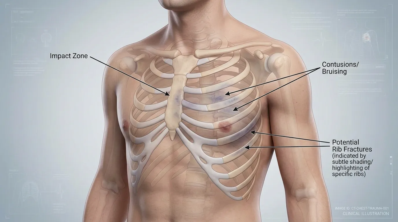
The bone has three parts. The manubrium is the wide, handle-like top portion where the collarbone and first set of ribs attach, Cleveland Clinic explains. Below it, the body is the long, flat center of the bone, where the third through seventh sets of ribs attach. The xiphoid process at the bottom is a small, pointed tip made mostly of cartilage, which begins to calcify and turn to bone with age.
The whole bone measures about 6 inches from top to bottom, by Cleveland Clinic‘s description. The sternum does not move the chest on its own, but the cartilage joining it to the ribs allows the minor motions that occur with every breath.
Belt Loading and Steering Wheel Impact
Sternal fractures most commonly result from blunt trauma to the front of the chest wall and from deceleration injuries, with a reported incidence of 3.0% to 6.8% in motor vehicle collisions, according to a 2022 editorial in Radiologia Brasileira. In a National Trauma Data Bank review the editorial cites, motor vehicle collisions were the leading mechanism at 68% of patients, followed by falls and motorcycle accidents at 7.9% each.
The force typically arrives as a direct blow to the front of the chest or as forced deceleration, the editorial explains. StatPearls is more specific about the car: sternal fractures typically result from the chest striking the steering wheel, with most injuries occurring in older vehicles lacking airbag deployment.
Shoulder belts changed the numbers. Seat belt legislation requiring shoulder restraints has led to an increased incidence of sternal fractures, the Radiologia Brasileira editorial states. The page on seatbelt injuries covers the wider set of injuries a belt can leave behind.
Older Occupants and Bone Strength
Sternal fractures are more common in older patients, which appears to be due to the elastic chest wall of younger patients, according to StatPearls. The same review names patients on long-term steroid therapy, postmenopausal women, and older patients as being at increased risk.
StatPearls describes sternal insufficiency fractures, which frequently correlate with pronounced thoracic kyphosis resulting from multiple thoracic compression fractures, especially in advanced osteoporosis.
Symptoms in the First Hours and Days
Pain sits at the front of the chest, over the bone. Sternal fractures typically present with anterior chest wall pain that deep breathing and coughing can make worse, and up to one-fifth of cases may also have shortness of breath, according to StatPearls.
Point tenderness over the sternum is common. Approximately half of reported cases may also present with soft tissue swelling, bruising, or a deformity that can be felt, StatPearls notes, and fracture-related crepitus may be noticeable when the area is pressed. Inspection of the front of the chest may show bruising with or without a step deformity, according to a 2013 review in the Journal of Emergencies, Trauma and Shock.
Some symptoms point past the bone. The card below draws on two MedlinePlus pages: the symptoms it lists for a bruised heart muscle, and the reasons its rib fracture aftercare page gives for contacting a provider.
Chest Injury Warning Signs
- Pain that does not allow deep breathing or coughing despite pain relievers
- Shortness of breath
- A cough or more mucus, especially if it is bloody
- A fever
- A feeling that the heart is racing
- Lightheadedness or weakness
- Nausea or vomiting
Fracture Locations Along the Sternum
Most sternal fractures are transverse, and any displacement occurs in the sagittal plane, according to the Radiologia Brasileira editorial.
The editorial summarizes a cohort of 108 patients in which the fracture was only in the manubrium in 59.3%, only in the body of the sternum in 38.0%, and in both places in 2.7%. Morbidity rates were higher with manubrium fractures than with fractures of the body, as was the incidence of accompanying bone fractures and organ injuries, and the cohort’s authors stated that a manubrium fracture can indicate the severity of trauma and a poor prognosis.
In that cohort, mortality was significantly higher among patients with comminuted fractures than among those with other fracture types, the editorial reports.
Why Kentucky Hires
Sam Aguiar Injury Lawyers
Heart Bruising and Associated Trauma
Car accidents are among the most common causes of myocardial contusion, a bruise of the heart muscle, according to MedlinePlus, which lists them first and says a severe contusion may lead to signs and symptoms of a heart attack.
Isolated sternal fractures are rarely associated with blunt cardiac injury, and the degree of fracture displacement is not necessarily associated with blunt cardiac injury either, according to the Radiologia Brasileira editorial.
In a review of 1,867 patient records in the National Israeli Trauma Registry, cited in the same editorial, none of the patients with an isolated sternal fracture needed a breathing tube, a chest tube, or a thoracotomy, compared with 16.9% of the polytrauma patients with sternal fractures.
Chest wall injuries include rib fractures, flail chest, and sternoclavicular dislocation, and thoracic spine compression fractures, as well as trauma to the head, neck, and extremities, are also common, the Journal of Emergencies, Trauma and Shock review reports. The page on thoracic spine injuries covers the mid-back side of that pattern.
Three Chest Injuries Side by Side
A broken sternum, a broken rib, and a bruised heart muscle share a location and some symptoms. The table lines them up using StatPearls, the Journal of Emergencies, Trauma and Shock review, and the MedlinePlus rib fracture and myocardial contusion pages.
| Feature | Sternal Fracture | Rib Fracture | Myocardial Contusion |
|---|---|---|---|
| What is injured | The breastbone at the center of the chest | One or more rib bones | The heart muscle, bruised |
| Typical symptoms | Sternal pain worse with deep breathing and coughing; localized tenderness | Pain when breathing, coughing, or moving the upper body | Pain in the front of the ribs or breastbone; racing heart; shortness of breath |
| Common tests | Lateral chest x-ray; chest CT | Checked for other chest and organ injuries | ECG, troponin blood tests, echocardiogram |
| Usual course | Most isolated fractures heal over an average of 10 weeks | Healing takes at least 6 weeks | Mild cases usually recover completely |
X-Ray Views and CT Scans
A chest x-ray is usually the first imaging test when a sternal injury is suspected, according to the Radiologia Brasileira editorial. A front-to-back, or anteroposterior, view has been shown to be only 50% sensitive for sternal fractures. A lateral view taken from the side has greater sensitivity and is typically diagnostic, because most of these fractures are transverse.
The sources rank the tests differently. The Journal of Emergencies, Trauma and Shock review called a lateral chest radiograph the gold standard in 2013. The 2022 editorial says computed tomography (CT) continues to be the gold standard and has been shown to be superior to lateral x-ray, and it cites a large retrospective study of thoracic trauma in which 94% of sternal fractures were visible only on chest CT.
CT also looks beyond the bone. A CT scan detects associated thoracic injuries in over 80% of patients with traumatic sternal fracture, the editorial notes. Ultrasound is another option: StatPearls says ultrasonography can detect sternal fractures with equal or greater sensitivity than plain radiography.
Cardiac Testing in the Emergency Room
Electrocardiograms should be conducted to look for signs of myocardial contusion, such as arrhythmias, conduction disturbances, sinus tachycardia, or ST-segment changes, according to StatPearls.
Enzyme markers of cardiac injury may be helpful if a cardiac contusion is suspected, with troponin elevation particularly indicative of heart muscle injury, StatPearls notes. Echocardiography gives a direct view of wall motion abnormalities.
Observation follows when a heart bruise is found. In most cases the patient is closely monitored for at least 24 hours, with an ECG done continually to check the heartbeat and heart function, according to MedlinePlus.
Stable patients with isolated sternal fractures, a normal ECG, and normal cardiac enzymes are expected to have a benign course and can be discharged home with pain medication and follow-up within the first 24 hours, the Journal of Emergencies, Trauma and Shock review states.
Pain Control and Surgical Fixation
Adequate pain control is the mainstay of treatment for isolated sternal fractures, according to StatPearls. Hospital admission is unnecessary if the clinical evaluation identifies no concerns, while patients with associated injuries inside the chest, hemodynamic instability, or uncontrolled pain should be admitted for observation.
Isolated and stable sternal fractures can typically be managed without surgery, while unstable fractures often need surgical fixation, StatPearls explains. Fixation may also be required for significantly displaced fractures and in rare cases of chronic sternal pain or nonunion. Surgeons can perform it with sternal wires or with plates and screws, according to the Journal of Emergencies, Trauma and Shock review.
For rib fractures, MedlinePlus describes slow deep-breathing and gentle coughing exercises every 2 hours to help prevent a collapsed lung or pneumonia, sometimes with a device called an incentive spirometer. The same page explains that patients do not get a binder or bandage around the chest, because it would keep the ribs from moving with breathing and coughing.
Healing Time and Return to Daily Activity
Most isolated sternal fractures heal on their own over an average period of 10 weeks, according to StatPearls, which describes an excellent prognosis for isolated fractures with an overall mortality rate of 0.7%. The Journal of Emergencies, Trauma and Shock review puts the average time to complete recovery at 10.4 weeks.
Chest pain can persist for 8 to 12 weeks after the injury, StatPearls notes. Advanced age, osteoporosis, prolonged steroid use, and diabetes raise the risk of delayed healing, the same review adds.
The MedlinePlus rib fracture aftercare page stresses staying active instead of resting in bed all day and avoiding movements that put painful pressure on the injured area, such as crunches and pushing, pulling, or lifting heavy objects. Return to work depends on the type of job, the page notes, and a provider sets the timing for sports and other high-impact activity.
Lung Infections, Nonunion, and Lasting Pain
Painful inspiration may lead to atelectasis, pneumonia, and other pulmonary complications, according to StatPearls. The Journal of Emergencies, Trauma and Shock review draws the same line, saying apprehension due to pain can impair ventilation and predispose a patient to chest infection.
Nonunion, false joint formation, and overlap deformities of sternal fractures are rare but may require delayed surgery, StatPearls reports. The 2013 review lists non-union among the long-term complications and adds that a painful false joint, called a pseudarthrosis, may develop.
Rare sequelae of sternal fractures include osteomyelitis, sternal abscess, and mediastinitis, according to the Journal of Emergencies, Trauma and Shock review.
Serious heart injuries can increase the risk of heart failure or heart rhythm problems, MedlinePlus warns, though people with a mild myocardial contusion recover completely most of the time.
Records From the Emergency Chart Onward
A sternal fracture leaves records in several departments. The emergency chart holds the chest x-ray or CT, the ECG tracings, and any troponin results, and the admission or discharge note records why the patient stayed for observation or went home.
Specialist records arrive later, when they are needed. An echocardiogram report, a cardiology consult, a physical therapy plan, or an operative note for wires or plates each documents a different part of the injury, and each facility keeps its own copy of what it produced.
A dated log of pain levels, nights of lost sleep, missed workdays, and tasks that had to wait shows how long a chest injury limited ordinary life. Kept with the emergency imaging and the ECG reports, that log puts the whole course of a sternal fracture in one place.
Breastbone Still Aching Weeks After the Crash?
A broken sternum can mean a CT in the trauma bay, a night on a heart monitor, and weeks of pain with every breath or cough, and an insurer may judge the whole injury by a single imaging report. Sam Aguiar Injury Lawyers gathers the emergency imaging, the ECG and troponin results, the follow-up notes, and any cardiology or surgical records so each stage of your recovery is on paper. Under our Bigger Share Guarantee®, you always walk away with more than the firm after bills, liens, and case costs. Start with a free case review.