

Chest Injuries After a Car Accident

Key Takeaways
- Motor vehicle collisions are the leading cause of chest trauma, according to the StatPearls review of thoracic trauma, and pneumothorax occurs in 30% of chest trauma.
- Chest radiographs can only diagnose about 50% of isolated rib fractures, according to the StatPearls chapter on rib fractures, which calls chest CT the gold standard for detecting them.
- Signs of a bruised lung may not appear on a chest x-ray for 6 to 48 hours after the injury, according to the StatPearls chapter on pulmonary contusion, though CT can detect a contusion immediately after trauma.
Chest Wall and Thoracic Organs
The ribs protect the organs and structures of the thoracic cavity, and the StatPearls chapter on rib fractures counts 12 pairs of them. The first seven attach in front to the sternum and in back to the spinal column, the chapter explains, ribs 8 through 10 connect to the costal cartilage of the sternum, and ribs 11 and 12 are called floating ribs because they attach only in back.
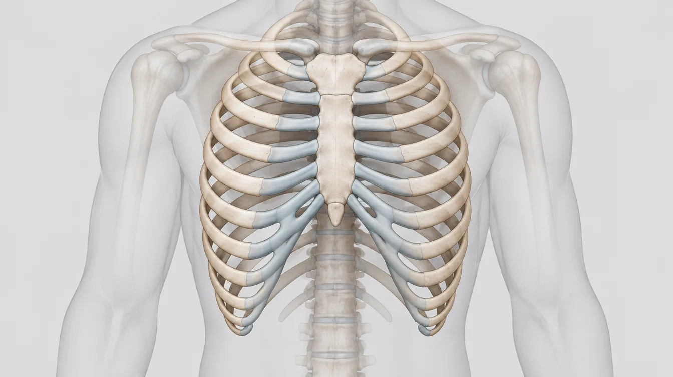
Inside that cage, the StatPearls review of thoracic trauma lists the great vessels as the aorta, pulmonary artery, superior and inferior vena cavae, and pulmonary veins, and it describes the trachea and bronchial tree carrying air from the larynx down to the alveoli, where gas exchange takes place. There is a space between each lung and the chest wall, and MedlinePlus explains that air escaping into it puts pressure on the lung.
An intercostal nerve, artery, and veins run beneath each rib, the StatPearls rib fracture chapter notes. The same chapter says any rib fracture should warrant a thorough evaluation for injury to the lungs, heart, kidney, spleen, liver, and nearby nerves and blood vessels.
Crash Forces and Injury Mechanisms
Motor vehicle collisions are the leading cause of chest trauma, according to the StatPearls thoracic trauma review, and most acute traumatic aortic injuries also come from crashes. The review calls blunt trauma a more common cause of traumatic injuries and says it may be life-threatening.
Trauma teams ask about the crash itself. Extensive vehicle damage, intrusion into the passenger space, prolonged extrication, airbag deployment, and steering wheel damage are risk factors for significant injury in a collision, the same review states, and the direction and magnitude of force must be documented.
Crushing or deceleration is the primary mechanism of cardiac injury, according to StatPearls, which describes the heart being compressed between the sternum and the thoracic spine, rapid deceleration, and increased blood return to the heart that overdistends a chamber to the point of rupture.
Rib Fractures and Flail Chest
A broken rib is a common injury, and Mayo Clinic names falls, car accidents, and contact sports as the most common causes. Many are simple cracks that hurt but are not typically dangerous, according to the same page. More serious breaks can split a rib into pieces, and Mayo Clinic says the sharp edges can injure the lungs, blood vessels, or other organs.
Possible complications depend on which ribs are broken, Mayo Clinic explains. In its breakdown, a sharp edge from a broken upper rib can tear a major blood vessel, including the aorta, a broken middle rib can puncture a lung and cause it to collapse, and broken lower ribs can damage the spleen, liver, or kidneys. Ribs 1 through 3 are the hardest to break and signify a significant degree of trauma if fractured, according to the StatPearls rib fracture chapter.
Flail chest is defined by fractures of 3 or more consecutive ribs in at least 2 places, leaving a free-floating segment of chest wall, according to the StatPearls chapter on flail chest. The chapter explains that the segment collapses inward during inspiration while the rest of the chest wall expands outward. It attributes approximately 75% of major trauma cases leading to flail chest to motor vehicle collisions, puts flail chest in roughly 7% of patients with chest trauma, and calls pulmonary contusion in the lung next to the flail segment nearly universal.
Symptoms That Appear After a Collision
The pain of a broken rib may get worse with a deep breath, with pressure on the injured area, or when bending or twisting, according to Mayo Clinic. A sternal fracture typically presents with pain in the front of the chest wall, and the StatPearls chapter on sternal fractures says up to one-fifth of patients may also be short of breath, with deep breathing and coughing able to make the pain worse.
A collapsed lung commonly causes sharp chest or shoulder pain made worse by a deep breath or a cough, MedlinePlus reports. A larger pneumothorax causes more severe symptoms, MedlinePlus adds, including chest tightness, lightheadedness and near fainting, cyanosis from lack of oxygen, and increased effort of breathing.
Symptoms of a pulmonary contusion can appear right after an injury or several hours to days later, according to Cleveland Clinic, and a person might notice slight changes, like a cough or breathlessness, before the symptoms become serious. On examination, the StatPearls thoracic trauma review directs clinicians to look for bruising and abrasions such as the seat belt sign, and it notes that chest wall contusions in a seatbelt pattern may produce point tenderness over the ribs.
Chest Warning Signs
- Trouble breathing, or pain with a deep breath after a crash
- Pressure, fullness, or squeezing pain in the center of the chest lasting more than a few minutes
- Chest pain that spreads to the shoulder or arm
- Sharp chest or shoulder pain that worsens with a cough
- Lightheadedness or near fainting
- Coughing up blood
- A rapid heart rate or rapid breathing
- A section of the chest wall that sinks inward with each breath in
Why Kentucky Hires
Sam Aguiar Injury Lawyers
Collapsed Lung and Blood in the Chest
MedlinePlus describes a collapsed lung as air escaping from the lung and filling the space between the lung and chest wall, where the buildup presses on the lung so it cannot expand as much as it normally does with a breath. The medical name is pneumothorax, and MedlinePlus lists rib fracture among the injuries that can cause it. Pneumothorax occurs in 30% of chest trauma, according to the StatPearls thoracic trauma review.
A simple pneumothorax can progress to a tension pneumothorax, the StatPearls review warns. In that form, the review explains, a flutter valve lets air flow one way into the pleural space and raises the pressure there. The review calls the diagnosis clinical: patients present with respiratory distress, rapid breathing, and low oxygen, and breath sounds are decreased or absent on the affected side.
Cleveland Clinic defines hemothorax as blood collecting in the pleural space, names an injury or trauma to the chest as the most common cause, and lists symptoms that may include shortness of breath, rapid breathing, a rapid heart rate, chest pain, low blood pressure, and restlessness. In blunt trauma, rib fractures may damage blood vessels inside the chest, and in severe cases a hemothorax can lead to hypovolemic shock if it is not addressed promptly, according to StatPearls.
Pulmonary Contusion in Blunt Trauma
A pulmonary contusion is a bruise on the lungs from a chest injury, and Cleveland Clinic says the most common cause is injury from a car accident. Cleveland Clinic explains that the injury damages blood vessels around the lungs, so blood and other fluids collect in the lung tissue.
Contusions are the most commonly identified lung injuries in blunt chest trauma, found in 30% to 75% of cases, according to the StatPearls chapter on pulmonary contusion. In mild injury, the chapter says, symptoms may develop gradually over 24 to 48 hours, and close monitoring is needed because a contusion can evolve even 24 to 48 hours after the trauma.
Pulmonary contusion should be suspected in patients with chest wall injury, especially if they present with low oxygen levels, the StatPearls thoracic trauma review advises. It adds that the degree of hypoxia often correlates with the size of the contusion and that a contusion may be silent during the initial evaluation.
Sternum Fractures and the Steering Wheel
Sternal fractures typically result from the chest striking the steering wheel, with most occurring in older vehicles that lack airbag deployment, according to the StatPearls chapter on sternal fractures. The chapter puts their incidence in motor vehicle collisions at 3% to 6.8% and notes that seat belt laws mandating shoulder restraints have, paradoxically, contributed to an increase in sternal fractures.
Morbidity from a traumatic sternal fracture typically depends more on its presentation and accompanying injuries than on the break itself, the StatPearls authors write, and up to two-thirds of sternal fractures involve associated injuries. Rib fractures, pulmonary contusions, and blunt myocardial injury are commonly associated with sternal fractures, according to the chapter.
Cardiac monitoring and pulse oximetry should be started for every emergency department patient with a sternal fracture, according to StatPearls, along with an electrocardiogram to look for signs of myocardial contusion such as arrhythmias, conduction disturbances, or ST-segment changes. The chapter adds that older patients warrant close monitoring because of a heightened risk of respiratory complications. The page on car accident sternum fractures covers breastbone injuries in more depth.
Heart and Aorta Injuries From Blunt Force
Blunt cardiac injury can range from minor myocardial contusions to cardiac rupture, valve damage, or pericardial tamponade, according to the StatPearls thoracic trauma review, and it often results from high-energy mechanisms such as motor vehicle collisions.
An electrocardiogram is a first-line test, yet StatPearls cautions that a normal ECG does not completely exclude significant cardiac trauma. Troponin levels may aid in diagnosing myocardial injury, the review says, and echocardiography is recommended for patients with abnormal ECG findings or hemodynamic instability to assess heart function, detect fluid around the heart, and identify structural damage.
StatPearls places most aortic injuries at the aortic isthmus, about 2 cm beyond the origin of the left subclavian artery, and says spiral CT angiography of the chest should be obtained after high-energy injury mechanisms to evaluate for aortic injury. The review describes thoracic aortic injuries as life-threatening and generally associated with poor outcomes.
Four Conditions Side by Side
The table sets four common crash injuries side by side, drawing on the sources cited on this page. They often overlap. The StatPearls thoracic trauma review says thoracic trauma rarely occurs in isolation, and the StatPearls flail chest chapter reports that flail chest occurs in isolation in fewer than 40% of cases.
| Feature | Rib Fracture | Sternal Fracture | Pneumothorax | Pulmonary Contusion |
|---|---|---|---|---|
| What is injured | A bone of the rib cage | The breastbone at the front of the chest | Air in the space between the lung and chest wall | Lung tissue, with blood and fluid collecting in it |
| Typical signs | Pain with deep breaths, pressing, bending, or twisting | Front chest wall pain, worse with deep breathing and coughing | Sharp chest or shoulder pain, worse with a breath or cough | Shortness of breath, coughing, coughing up blood |
| Chest x-ray | Finds about 50% of isolated fractures | Front view 50% sensitive, lateral view usually diagnostic | Small and medium ones may be missed on a film taken lying down | Signs may be delayed 6 to 48 hours |
| Common treatment | Pain relief, rest, ice, incentive spirometer | Pain relief; surgical fixation may be needed for significantly displaced or unstable breaks | Oxygen and rest if small, chest tube if large | Hospital monitoring, oxygen, pain medication |
| Usual healing | About six weeks for most | Average of 10 weeks when isolated | A small one may go away on its own | 5 to 7 days for most with supportive care |
Imaging With X-Rays, CT, and Ultrasound
A portable chest radiograph is required in most trauma codes, according to the StatPearls thoracic trauma review, yet the same review says small and medium pneumothoraces and hemothoraces may be missed on a chest film taken lying down. The StatPearls rib fracture chapter adds that chest radiographs can only diagnose about 50% of isolated rib fractures, and contusion signs may not appear on a chest x-ray for 6 to 48 hours after the injury, according to the StatPearls chapter on pulmonary contusion.
CT is more sensitive and specific than radiography or ultrasound for evaluating the structures in the middle of the chest, StatPearls states, and it is definitive in chest trauma, though the patient should be hemodynamically stable throughout the test. Chest CT is the gold standard for detecting rib fractures, according to the rib fracture chapter, although the fractures it detects may not be clinically significant.
The extended FAST ultrasound exam adds views of both lungs to look for pneumothorax and hemothorax, the thoracic trauma review explains, and the pulmonary contusion chapter calls ultrasound a vital bedside tool when a chest x-ray or CT cannot be obtained immediately. For the breastbone, a lateral x-ray view increases sensitivity and is usually diagnostic because most sternal fractures are transverse, according to the StatPearls sternal fracture chapter. The page on radiology imaging after a car accident explains how imaging reports are written and read.
Treatment From Pain Control to Chest Tubes
Pain management is critical in reducing respiratory complications after chest trauma, according to the StatPearls thoracic trauma review. Pain leads to splinting, which the review describes as involuntary guarding that can worsen or prevent healing, and it says splinting can lead to pneumonia in many cases. Mayo Clinic makes the same connection for broken ribs: good pain control helps a person breathe deeply, which lowers the risk of lung conditions such as pneumonia.
For simple, isolated rib fractures, the StatPearls rib fracture chapter describes conservative therapy with appropriate analgesia, rest, and ice, and it says an incentive spirometer should be encouraged to prevent pulmonary atelectasis and splinting. Pulmonary contusions have no specific treatment, Cleveland Clinic notes, so care focuses on monitoring in the hospital and keeping the condition stable with measures such as oxygen therapy, pain medication, and pulmonary hygiene.
Large pneumothoraces and hemothoraces call for prompt chest tube thoracostomy, according to StatPearls. A small pneumothorax may go away on its own over time with only oxygen treatment and rest, MedlinePlus says, while for a large one a chest tube is placed between the ribs and may be left in place for several days.
Most thoracic trauma cases can be managed without an operation, the thoracic trauma review notes. For severe injuries, particularly flail chest, surgical stabilization of rib fractures is recognized as a key treatment option, and early surgery within 48 to 72 hours is associated with less time on mechanical ventilation, shorter intensive care and hospital stays, and a lower incidence of pneumonia, according to the StatPearls flail chest chapter.
Recovery Ranges for Ribs, Sternum, and Lungs
Most broken ribs heal on their own in about six weeks, according to Mayo Clinic. Most isolated sternal fractures heal on their own over an average period of 10 weeks, the StatPearls sternal fracture chapter reports, though chest pain can persist for 8 to 12 weeks after the injury.
Most pulmonary contusions heal within 5 to 7 days with good supportive care, according to the StatPearls chapter on pulmonary contusion. Cleveland Clinic puts healing at about a week and says a longer hospital stay may be needed depending on severity.
The prognosis for isolated rib fractures is good, the StatPearls thoracic trauma review says, but lung or cardiac injury often leads to prolonged recovery. Older patients with sternal fractures may face prolonged recovery periods due to pain and osteoporotic bone, according to the sternal fracture chapter.
Pneumonia and Other Late Complications
Complications of chest trauma include pneumonia, acute respiratory distress syndrome, acute lung injury, mediastinitis, and arteriovenous and bronchopleural fistulas, according to the StatPearls thoracic trauma review. The review also reports that mortality increases by 19% and the pneumonia risk by 27% for each additional rib fractured.
Up to 20% of people with a pulmonary contusion develop pneumonia, Cleveland Clinic reports, and the StatPearls pulmonary contusion chapter lists pneumonia, ARDS, and respiratory failure among the possible results of a contusion. Retained blood within the pleural cavity may lead to infection, resulting in empyema or fibrothorax, a condition in which pleural scarring restricts lung expansion, according to StatPearls.
After flail chest, adverse events are common and often result in prolonged disability, with pain persisting for months or even years in some cases, according to the StatPearls flail chest chapter, which lists severe chronic pain, chest wall deformity, and loss of exercise endurance among the complications. Nonunion and false joint formation after a sternal fracture are rare, the sternal fracture chapter notes, and advanced age, osteoporosis, prolonged steroid use, and diabetes raise the risk of delayed healing.
Medical Records That Trace the Injury
The first record of a chest injury is usually the emergency chart. The StatPearls thoracic trauma review says the circumstances of the event, including the direction and magnitude of force, must be documented, and the exam notes that follow record findings such as point tenderness, crepitus, breath sounds, and any seat belt mark across the chest.
Later entries show how the injury behaved: repeat imaging, oxygen needs, chest tube placement and removal, pain management, and follow-up visits. Work and activity restrictions written into those notes show what the injury changed from one week to the next. The page on thoracic spine injuries covers the vertebrae and spinal structures behind the chest.
Chest symptoms that change over the first days after a crash are easier to follow when the emergency notes, each imaging report, and any chest tube or monitoring records are kept together. A dated log of breathing trouble, coughing, and pain with movement gives each new provider a baseline to compare against.
Chest Still Hurting Weeks After the Crash?
Broken ribs, a bruised lung, or a cracked sternum can keep every breath and cough painful long after the emergency room visit, and an insurer reading the chart may still call the injury minor. Sam Aguiar Injury Lawyers lines up the emergency notes, the CT reports, and the follow-up records so the full course of your chest injury is on paper. Under our Bigger Share Guarantee®, you always walk away with more than the firm after bills, liens, and case costs. Start with a free case review.